The World Health Organization defines a burn as an injury to the skin or other organic tissue by heat, radiation, radioactivity, ultraviolet radiation, electricity, friction or contact with chemicals. Frostbite is also considered a thermal burn
Skin injuries as well as respiratory damage from smoke inhalation are also considered to be burns.
HOW ARE BURNS CLASSIFIED?
|
-
PARTIAL THICKNESS (1ST DEGREE)
- superficial burns are limited in depth to the first 2 or 3 of the 5 layers of the epidermis
- characterized by erythema, hyperemia, tenderness and pain
- no vesicles or blisters
- due to the shallow depth, regeneration of skin will occur within several days
- burns involve entire epidermis (blue zone) and upper third of dermis (mauve zone)
- skin will usually appear red
- injury to microvessels that perfuse the skin results in wet wounds with bullae and blisters
- very painful; pain increases when exposed to air currents
- will heal within 1 - 6 weeks with minimal scarring
Assessment findings: Superficial Partial-Thickness
-
-
PARTIAL THICKNESS (2ND DEGREE)
- SUPERFICIAL PARTIAL-THICKNESS
-
- tenderness
- blanches with pressure
- wet, weeping, erythematous skin
- clear blisters
- usually pink color, but may be white
- brisk bleeding with 21 gauge needle pinprick
DEEP PARTIAL THICKNESS
- burn includes the entire epidermis and deep into the dermis
- impairment of blood supply often limits fluid leakage
- will heal spontaneously by slow granulation, often leaving unstable epithelium, scarring and contractures
- grafting is the desired treatment as it improves the quality of the healing (better cosmetic and functional healing) and reduces the opportunity for infection can progress to full thickness injuries if infection develops
Assessment findings: Deep Partial Thickness
- no sensation to touch or dull sensation
- white or fixed red coloration
- non-blanching with pressure
- blisters may be present
- delayed bleeding with 21 gauge needle prick
-
- blisters are not usually present and only a modest amount of plasma leakage appears on the wound
- wound usually red with white appearance in center
- dermal necrosis and surface protein gives burn surface a yellow appearance
Full Thickness Burn (3rd degree)
- results in destruction of skin through all layers of epidermis and dermis, extending into subcutaneous fat and underlying tissue
- subcutaneous layer contains hair follicles and sweat glands and is poorly vascularized; this is below the stratum germinativum layer (dark blue area) which is responsible for the generation of new skin cells
Assessment findings: Full Thickness Burn
- burn appears white, red or brown and is often charred and leathery in appearance
- skin is usually dry and insensitive to pin or palpation
- no blanching with pressure, or capillary refill or bleeding with pinprick
- if skin is broken, subcutaneous fat may be visible
- associated with extensive fluid and electrolyte imbalances, altered thermoregulation, metabolic disturbances and infection
- although small wounds will contract and eventually heal, larger wounds require grafting
-
Fourth and Fifth Degree Burn
- Burns that are deep below subcunatneous tissues that include fascia and muscle layers are referred to as fourth degree burns.
- A fifth degree burn is one that requires amputation or loss of body parts.
-
|
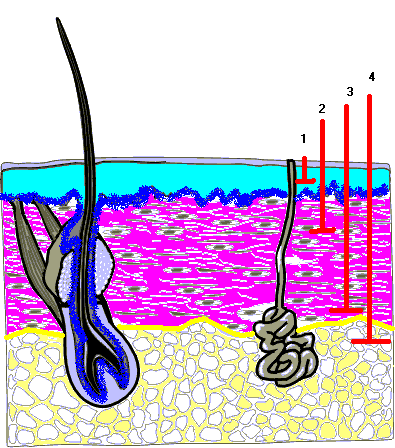
Diagram 1
Epidermis:
blue area
Stratum Germinativum:
dark blue area
Dermis:
mauve area
Subcutaneous Fat:
yellow area
Burn Depths:
partial thickness (1)
moderate partial thickness (2)
deep partial thickness (3)
full thickness (4)
Diagram 1
|
Calculation of Percentage of Burned Area
Management and Monitoring of the Burned Patient
Brenda Morgan. (October 23, 2000)
Last Update: January 15, 2019
References:
Buck, D., Fedorowicz, Z., and Ehrlich, A. (2018). Major burns. DynaMed updated October 23, 2018.
http://web.a.ebscohost.com.proxy1.lib.uwo.ca/dynamed/detail?vid=2&sid=af18d660-cd94-4141-84d3-54691187c6f0%40sessionmgr4007&bdata=JnNpdGU9ZHluYW1lZC1saXZlJnNjb3BlPXNpdGU%3d#AN=113804&db=dme
World Health Organization https://www.who.int/violence_injury_prevention/other_injury/burns/en/
Classification of Burns. https://www.urmc.rochester.edu/encyclopedia/content.aspx?ContentTypeID=90&ContentID=P09575
Burn classification - burns are acute injuries. White Paper. American Burn Association.