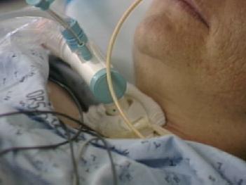
TRACHEOSTOMY
A tracheostomy is a hole in the neck that is created just below the Adam's Apple. It punctures the trachea (or windpipe) and is created in order to insert a Tracheostomy Tube. A tracheostomy tube can be used to deliver mechanical breaths. It is also used to reduce the chance that a patient will choke on their own saliva and develop pneumonia.
Tracheostomy tubes are more comfortable for patients who require prolonged mechanical ventilation than tubes inserted through the nose or mouth (endotracheal). Tracheostomy tubes are shorter than endotracheal tubes. The shorter tube can allow more air to reach the lungs and make breathing easier for patients with very severe lung disease.
The term percutaneous (through the skin) describes the most common method for inserting a tracheostomy tube in the critical care unit. This is done at the bedside under anaesthetic by a specialy trained surgeon. A bronchoscope is used to look at the inside of the trachea while the tracheostomy is being done. Occasionally, tracheostomies are performed in the operating room through a small incision in the neck.
The top of the windpipe has a thin piece of cartlidge or tissue called the epiglottis. The epiglottis normally closes when we swallow to prevent saliva and food from entering our lungs. When a patient has a breathing tube inserted through the nose or mouth (endotracheal tube), the epiglottis is kept in a permanently open position. This allows saliva to enter the lungs (called aspiration).
All breathing tubes have a donut shaped balloon located around the outside of the tube (like a life preserver), called a cuff. When the cuff is inflated, air can only enter and leave the lungs through the inside of the tube. When a tube is inserted through the epiglottis (such as an endotracheal tube), the cuff must be inflated at all times to prevent aspiration of saliva. If the patient is on a mechanical ventilator, inflation of the cuff also keeps the mechanical breath from leaking out around the tube, keeping the air in the lung.
Speech is produced when exhaled air passes through the vocal cords, causing them to vibrate. The vocal cords are located at the top of the trachea near the epiglottis. If the cuff of a breathing tube is inflated, exhaled air will leave through the inside of the tube, and never reach the vocal cords. Thus, patients cannot create sound or speech when they have a breathing tube in place with an inflated cuff.
Tracheostomy tubes are inserted below the vocal cords, and also have inflatable cuffs. Because a tracheostomy tube is below the vocal cords and epiglottis, the cuff can be deflated if the patient is able to control the opening and closing of their epiglottis when they swallow (or able to "gag"). If the patient with a tracheostomy requires mechanical ventilation, the cuff will usually need to be inflated to prevent loss of the mechanical breath around the tube. As long as the cuff needs to be inflated, speech is not possible.
Patients requiring long term ventilation, who are able to control the opening and closing of their epiglottis, may be able to tolerate periods with the cuff deflated, allowing speech.
Reviewed: October 30, 2018