Question
of the Week: December
3, 1999
|
How
are burns classified?
|
| How
are Burns Classified? |
|
Partial Thickness
(1st
degree)
-
superficial
burns are limited in depth to the first 2 or 3 of the 5 layers of the epidermis
-
characterized
by erythema, hyperemia, tenderness and pain
-
no
vesicles or blisters
-
due
to the shallow depth, regeneration of skin will occur within several days
Moderate Partial
Thickness
(2nd
degree)
-
burns
involve entire epidermis (blue zone) and upper third of dermis (mauve zone)
-
skin
will usually appear red
-
injury
to microvessels that perfuse the skin results in wet wounds with bullae
and blisters
-
very
painful; pain increases when exposed to air currents
-
will
heal within 1 - 6 weeks with minimal scarring
Deep Partial
Thickness
(2nd
degree)
-
burn
includes the entire epidermus and deep into the dermis
-
impairment
of blood supply often limits fluid leakage; blisters are not usually present
and only a modest amount of plasma leakage appears on the wound
-
wound
usually red with white appearance in center; blanches following assessment
for capillary refill
-
dermal
necrosis and surface protein turns gives burn surface a yellow appearance
-
will
heal spontaneously by slow granulation, often leaving unstable epithelium,
scarring and contractures
-
grafting
is the desired treatment as it improves the quality of the healing (better
cosmetic and functional healing) and reduces the opportunity for infection
can
progress to full thickness injuries if infection develops
|
Full Thickness
Burn
(3rd
degree)
-
results
in destruction of skin through all layers of epidermis and dermis, extending
into subcutaneous fat and underlying tissue
-
subcutaneous
layer contains hair follicles and sweat glands and is poorly vascularized;
this is below the stratum germinativum layer (dark blue area) which is
responsible for the generation of new skin cells
-
burn
appears white, red or brown and is often charred and leathery in appearance
-
skin
is usually dry and insensitive to pin or palpation
-
if
skin is broken, subcutaneous fat may be visible
-
associated
with extensive fluid and electrolyte imbalances, altered thermoregulation,
metabolic disturbances and infection
although
small wounds will contract and eventually heal, larger wounds require grafting
|
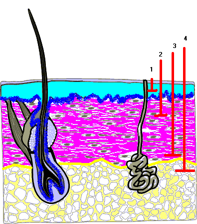
| Diagram
1
Epidermis:
blue
area
Stratum
Germinativum:
dark
blue area
Dermis:
mauve
area
Subcutaneous
Fat:
yellow
area
Burn
Depths:
partial
thickness (1)
moderate
partial thickness (2)
deep
partial thickness (3)
full
thickness (4)
|
|
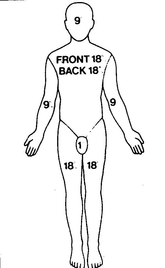
| How
is the "Percentage of Burned Area" Calculated? |
|
| Estimation
of the size of the burn is done using the "rule of nines" shown in Diagram
2. The corresponding area of burn is identified on the diagram, and
the total percentage is calculated as the sum of the burned areas.
The
type or thickness of the burn can also be recorded on the diagram.
Many burns are mixed in nature, for example, the outer edges may be partial
thickness, while areas of deep partial and full thickness burns may extend
toward the center. The appearance of the burn, along with the presence
of blanching, blisters and pain helps to determine the extent of the injury.
The
percentage of burns may change as the tissue injury evolves and more extensive
tissue damage becomes evident. Infection can increase the severity
of the burn over the course of the injury.
|
Diagram
2
|
Brenda Morgan. (December
1, 1999)
References:
Thelan,
L., Urden, L., Lough, M., & Stacy, K. Critical Care Nursing: Diagnosis
and Management. Mosby: Toronto. pp. 1141-1170.