Question
of the Week:
|
|
Answer:
- broad QRS > .11 seconds
- rSR' pattern in V1
- broad terminal S wave in Leads I, aVL and V6
- there may be secondary T wave changes (inversion)
Normally, the right and left ventricles depolarize at the same time. This results in the depolarization of both ventricles within .12 seconds (a narrow QRS). If one bundle branch is blocked, the ventricle with the normal bundle branch will depolarize first. The ventricle with the diseased bundle branch will depolarize after the normal ventricle. This prolonged time for depolarization of both ventricles produces a broad QRS. In leads that have the best view of the left or right ventricle, the asymmetrical depolarization can be seen by a change to the normal pattern.
Although both ventricles normally depolarize together, the left ventricle sends a stronger electrical signal. The sum of the forces from the left ventricle are greater than from the right, therefore, the right ventricular depolarization is usually "hidden" under the stronger left ventricular depolarization wave.
In right bundle branch block, the left ventricle depolarizes first. The right ventricle will depolarize after the left.
Right Ventricular
Lead
The V1
lead
is closest to the right ventricle. It will identify depolarization of the
right ventricle (current flowing towards the lead) as a positive
deflection and left ventricular depolarization (current flowing away from
the lead) as a negative deflection. In normal depolarization, the
septum depolarizes before the bundle branches. The septum depolarizes
from left towards right.
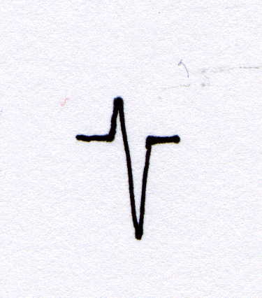
Normal V1
In a normal V1
pattern, the QRS begins with a small initial "r" wave, indicating depolarization
of the septum towards the lead. This is followed by simultaneous
transmission of the impulse through the right and left bundle branches,
producing simultaneous left and right ventricular depolarization.
Because the left ventricle sends a stronger impulse, the net sum of the
ventricular forces is away from V1, producing a downward
deflection (or S). The right ventricular depolarization wave (upward
deflection) is buried in the S wave. Thus, the normal QRS pattern in V1
is rS.
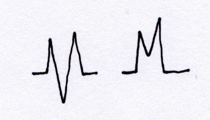
V1
in Right Bundle Branch Block (RBBB)
In RBBB, the
septum continues to depolarizes first, producing a small initial "r" in
V1. Because the right bundle is blocked, the
impulse then travels towards the left bundle branch only, producing a downward
deflection or S wave. Following depolarization of the left ventricle,
the right ventricle depolarizes from a "back door" impulse, producing a
second R wave in V1 (a second R wave is called "R
Prime" and is identified as R'). This causes the characteristic rSR'
in V1 of RBBB. Because depolarization is abnormal,
abnormal repolarization may also be seen by T wave changes.
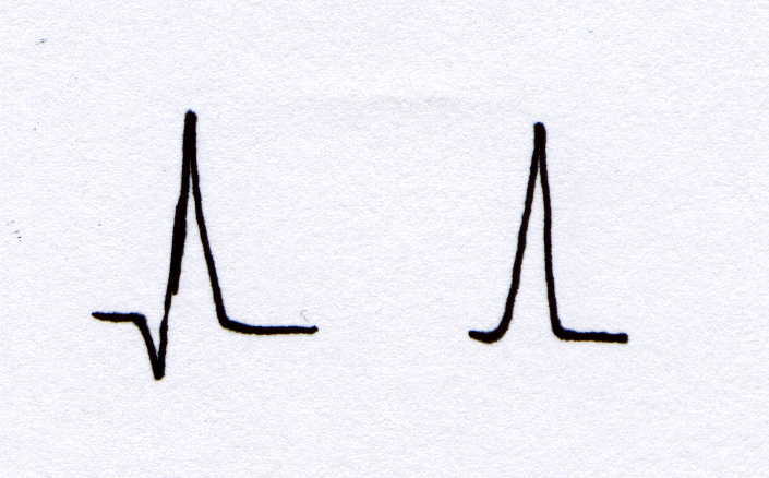
Normal I, aVL
and V6
Leads I, aVL
and V6 are in the best position to observe "leftward"
depolarization. A small initial "q" wave may be present normally,
reflecting the initial depolarization of the septum from left to right
(or away from I, aVL and V6).
The right and left ventricles then depolarize simultaneously. Because
both ventricles depolarize within the same period of time, the QRS is narrow
(< .12 seconds). Because the left ventricular force is strongest,
leads I, aVL and V6 display a
tall "R" wave. The right ventricular depolarization wave (away
from I, aVL and V6) is buried
in the R wave. The normal pattern is a narrow "R" or narrow qR pattern.
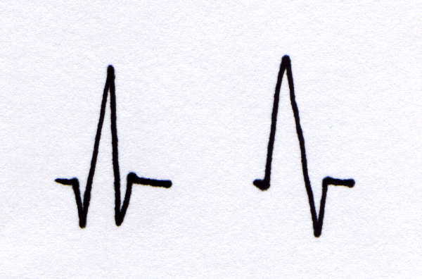
Leads I, aVL
and V6 in Right Bundle Branch Block (RBBB)
With RBBB, the
right ventricle depolarizes after the left. This produces a wide
QRS with a terminal S wave in I, aVL and V6.
The QRS pattern in I, aVL and V6 appears
as a qRS or RS pattern.
| Normal V1 Appearance | V1 Appearance in RBBB |
|
|
|
|
| Normal I, aVL and V6 Appearance | Leads I, aVL and V6 in RBBB |
|
|
|
|
References:
Conover, M. (1990). Pocket Guide to Electrocardiography. Mosby: Toronto.
Conover, M. (1984). Understanding Electocardiography (4th Edition). Mosby: Toronto.
Brenda Morgan
Clinical Educator, CCTC
February 27, 2000
|
|