|
BRAIN INJURY; HEAD INJURY A brain injury refers to damage to the brain. Head Injury is another name for brain injury. If a specific area of the brain is damaged the patient will usually experience difficulties performing functions that are controlled by the damaged area of the brain. For example, they may have problem moving or feeling a part of their body. They might have problems with speech or comprehension or they may have problems with memory (Image 1). Diffuse Axonal Injury is a term used to describe a very serious type of brain injury. Diffuse Axonal Injury usually occurs when a patient has been involved in an accident where he or she came to a rapid and abrupt stop (called a "rapid deceleration injury). The brain is surrounded by a layer of fluid. In a rapid deceleration injury, the brain will be forced forward into the skull bone. It will then bounce backwards and twist back and forth until it comes to a stop. The injury is "spread all over" (diffuse), and causes shearing and tearing of the axons (nerves). This type of injury is not always visible when brain imaging tests are done right after the accident, but often cause very severe disabilities. The brain is surrounded by 3 very thin protective membranes called the meninges. The outer layer that is located immediately below the skull bone is called the Dura. Below the dura is a membrane called the arachnoid membrane. The membrane closest to the brain is called the pia (Image 2). After injury or damage to a blood vessel, patients can bleed in between these layers. Bleeding between the dura and bone is called an epidural hematoma ("epidural" means above the dura; "hematoma" means blood clot). This type of bleeding is usually from an artery and can increase in size very quickly. We may watch a very small epidural hematoma and allow the patient to recover on their own, however, larger or rapidly growing hematomas may need urgent drainage by a neurosurgeon. If the bleeding increases, the hematoma will push against the brain and cause brain swelling and injury. Drainage or removal of the hematoma is called "evacuation". Patients can also have bleeding between the dura and arachnoid membrane. Bleeding in this space is called a subdural hematoma ("subdural" means below the dura; "hematoma" means blood clot). Bleeding in this space is usually from a vein, and may be slower to collect than an epidural bleed. Sometimes patients who are prone to hitting their head may be admitted with neurological changes, and found to have several old areas of bleeding. This is called "chronic subdural hematoma ("chronic" means long standing). Subdural bleeds can also be large and life-threatening and may also require surgery if they begin to press on the brain. The space between the arachnoid and pia membranes is the largest space. It is called the subarachnoid space (below the arachnoid membrane). It is filled with cerebral spinal fluid and contains the major arteries that supply the brain with oxygen. Bleeding can occur in the this space after trauma. Because the large arteries are situated within this space, a ruptured cerebral aneurysm (aneurysm in the brain) is the most common reason for a subarachnoid hemorrhage (bleeding in the subarachnoid space). When an area of the brain has been permanently damaged, the patient has experienced a Stroke. The brain can be injured for many reasons, including: trauma, lack of oxygen, bleeding, infection, toxins or blood clot. |
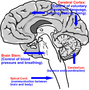
Image 1: The Brain |
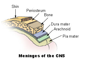
Image 2: Meningeal layers that cover the brain |
|
|
|
|
|
|
|
Last Reviewed: October 23, 2014
|
|