Sound Transmission in the Normal-Hearing Ear
Outer Ear
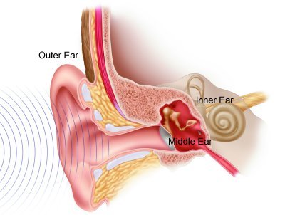
Sound is collected by the outer ear (also known as the pinnae). It travels down the ear canal and vibrates the eardrum. This turns an invisible energy (the acoustic signal) into visible movement of the eardrum (mechanical force).
Middle Ear
The eardrum movement causes movement of the middle ear bones. This bone movement provides an increase in the force of the energy as it moves from air-filled middle ear space to a fluid-filled cochlea.
Inner Ear
The cochlea is snail-shaped and is filled with tens of thousands of hair cells. The hair cells are embedded in structures that move in response to the waves created by the incoming sound. The movement of these hair cells results in a small electrical charge. This charge is sent by the auditory nerve up to the brain for interpretation as sound. The cochlea is arranged "tonotopically" like a piano keyboard. High frequencies are processed at the base of the cochlea (closest to the middle ear space), low frequencies are processed at the apex of the cochlea.
Hearing-Impairment
Most individuals with a severe to profound sensorineural hearing loss have damage to the hair cells that process sound (see illustration above). Because there are so few remaining hair cells, sounds must be very loud in order to just be heard. Like a piano that is missing "keys", the speech message may be unclear.
For some individuals with a hearing loss, a hearing aid can assist in the perception of sound. A hearing aid provides additional loudness to stimulate the remaining hair cells. As noted above, individuals with a severe to profound hearing loss have very few remaining hair cells limiting the benefit of a traditional hearing aid. The alternative for these individuals is a cochlear implant.


