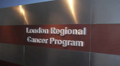
![]() |
About Us | ![]() |
Patients, Families & Visitors | ![]() |
For Health Professionals | ![]() |
Careers | ![]() |
Research & Training | ![]() |
Ways to Give | ![]() |
|---|
Scientist: London Regional Cancer Program, London Health Sciences Centre, London, Ontario
Robert Hardie Chair in Translational Prostate Cancer Research: Information
Director: Translational Prostate Cancer Research Group
Assistant Professor: Department of Surgery, Division of Urologic Surgery, University of Western Ontario, London, Ontario
Assistant Professor: Department of Oncology, University of Western Ontario, London, Ontario
Cross Appointment: Medical Biophysics
John Lewis, Ph.D.
Department of Oncology, University of Alberta
5-142C Katz Group Building
114th St and 87th Ave
Edmonton, AB T6G 2E1 CANADA
Office: 780-492-6113
Email: jdlewis@ualberta.ca
Non-invasive imaging and targeting of angiogenesis and metastasis using multivalent nanoparticles
Cancer begins when a cell begins dividing uncontrollably, eventually forming a visible mass or primary tumour. The ability of a primary tumour to grow beyond a few millimetres in size depends on its ability to stimulate angiogenesis. Without angiogenesis, a tumour is far more likely to remain dormant or regress. Thus, tumour dependency on neovascularization provides novel opportunities for the early detection and treatment of cancer. In urinary cancers, such as renal cell cancer, prostate cancer, and bladder cancer, advanced disease is a good candidate for antiangiogenic therapy because of its resistance to ordinary chemotherapy, radiotherapy, and hormonal therapy. Since current human antiangiogenic therapies have shown minimal effect on patient survival, it is critical that new targets be identified. Non-invasive strategies aimed at these targets have the potential to both specifically detect and treat solid tumours without biopsy or surgery and should lead to lower costs and higher quality of life. Thus, one of the primary interests in my laboratory is the development of nanoparticle-based imaging and therapeutic agents targeted at promising angiogenic factors.
Cells from the primary tumour begin to move and ultimately end up elsewhere in the body, where they then grow into secondary tumours. This process, called metastasis, is a complex multi-step process that has a significant impact on cancer mortality. Metastases are the cause of 90% of human cancer deaths. For prostate cancer, which is the most prevalent solid malignancy in Canadian men, the ten year survival for localized prostate cancer was recently reported at 73.1%. This survival rate drops precipitously to 6.81% for metastatic disease. Even when a tumour has not metastasized to a distant organ, movement of cells in a tumour can prove lethal because it can make complete surgical removal of the disease very difficult. We are therefore very interested in the study of the tumour microenvironment, with particular focus on the pathways and mechanisms that enhance tumour cell motility and metastasis.
Non-invasive imaging using multivalent nanoparticles: We recently demonstrated that multivalent viral nanoparticles are well suited for long term intravital vascular imaging (Nat Med 12, 354-360), and more recently have been using targeted trifunctional nanoparticles for the optical imaging of VEGF receptors on activated endothelial cells. The sensitivity and specificity of these viral nanoparticles has encouraged us to utilize multivalent trifunctional viral nanoparticles targeted towards angiogenic vasculature to facilitate the early detection and treatment of neoplasias and micrometastases. To this end, we are working to identify new targets of angiogenic blood vessels and to visualize the localization of these targets in vivo in animal models of cancer.

Fluorescent viral nanoparticles light up new blood vessels radiating from an epithelial carcinoma
Lewis JD, Destito G, Zijlstra A, Gonzalez MJ, Quigley J, Manchester MA, and Stuhlmann H (2006) Viral nanoparticles (VNPs) as tools for intravital vascular imaging. Nat Med 12:354-360.
Intravital real-time imaging of tumour cell invasion and metastasis: In order for haematogenous (through the bloodstream) metastasis to take place, tumour cells have to enter the vasculature. Cells from highly metastatic disease such as advanced prostate cancer disseminate efficiently in part because they intravasate (enter blood vessels) in high numbers. Intravasation has long been recognized as a rate-limiting step in metastasis. We have recently determined that intravasation is migration dependent, and that by inhibiting migration, one can block metastasis (Cancer Cell 13, 221-234). Nevertheless, the exact route by which intravasation takes place is poorly understood. Furthermore, while angiogenesis is clearly a requirement for metastasis to take place, it has not been determined whether metastatic cells enter new vasculature or larger, more established blood vessels. We have recently developed a comprehensive platform to quantitate the rate-limiting steps of metastasis and to visualize tumour cell behaviour in vivo using fluorescence intravital microscopy. Using quantitative assays and long term timelapse intravital imaging, we would like to determine the route of intravasation utilized by primary tumour cells as they metastasize.

Tumour cells invading the tissues surrounding a primary tumour are observed migrating along blood vessels
Zijlstra A, Lewis JD, Degryse B, Stuhlmann H and Quigley JP (2008) The inhibition of tumor cell intravasation and subsequent metastasis via regulation of in vivo tumor cell motility by the tetraspanin CD151. Cancer Cell 13:221-234.
Post-doctoral fellows
Hon Leong
Bhavik Manocha
Desmond Pink
Graduate Students
Choi-Fong Cho
Rae Lynn Nesbitt
Amy Robertson
Catalina Vasquez
Navid Baktash
Laboratory Manager
Amber Ablack
Research Associates
Alexandra Meilutis
Kelsey Taylor
Clinical Fellows (Uro-Oncology)
Ana Maria Autran
Ali Alzahrani
Vladimir Yutkin