There are a range of diagnostic tests used by clinicians:
Electroencephalogram (EEG)
The EEG or Electroencephalogram (pronounced ‘ee-lek-tro-en-seh-fah-lo-gram’) is a harmless diagnostic test that picks up electrical impulses from the brain and records them in a series of wavy lines. This test is a primary diagnostic tool for epilepsy.
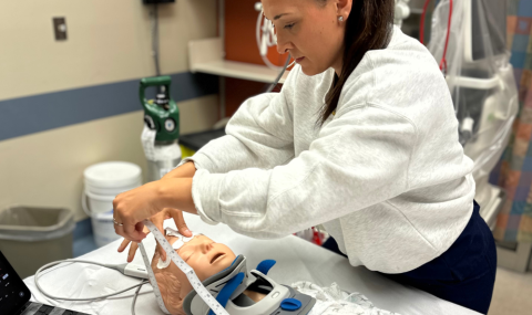
To perform an EEG, electrodes are placed on the scalp using a thick paste (conductive cream). A gritty material is used to prep the scalp to eliminate natural scalp oils so this may feel slightly scratchy.
A measuring tape and coloured crayon/marker may be used to locate the correct electrode placement (23 electrodes in total). Two additional electrodes will be placed on the patient's chest to monitor their heart.
The patient will feel nothing during the recording. Ideally, she/he will be calm and relaxed, but we recognize this is not always the case. The EEG will be carried out with your eyes open and your eyes closed. It is beneficial to record sleep if the patient happens to doze off as sometimes abnormalities are seen during sleep that is not present in wakefulness.
Hyperventilation (deep breathing) will be attempted for three minutes. Also, a flashing light will be placed in front of the patient. The entire EEG is video recorded to help with diagnosis.
Electrodes are removed easily. Electrode paste will be cleaned from the patient's hair, but some residue will remain. It is wise to bring a comb or brush to tidy up afterwards.
Several different types of EEGs may be ordered, including a routine EEGs (awake or asleep) which take around 25-30 minutes or a three-hour prolonged EEGs.
SPECT Scan
Single Photon Emission Computed Tomography (SPECT) scan is a type of nuclear imaging test that shows how radioactive tracers flow to tissues and organs. In epilepsy, this test is used to look at how the blood flows through the brain. The SPECT scan shows changes in blood flow during or in-between a seizure. This information can show doctors information about the location of seizures and whether they are contained in certain areas of the brain.
A SPECT scan takes an image of the distribution of a radioactive material (tracer). The tracer is what allows the doctor to see how the blood flows and visualize any accumulation.
There are two types of SPECT scans:
- Ictal SPECT scans occur during the epileptic seizure. The image on the left shows blood flow during a seizure and the one on the right shows blood flood between seizures.
- Inter-Ictal SPECT scans occur between seizures.
Procedure:
Patients who are potential epilepsy surgical candidates may require this testing for further information to see where the seizures are coming from in the brain. Epileptologists will order this test when the patient is admitted to the EMU. This procedure needs to be completed within four hours as the tracer, once drawn up is only good for five hours. Once a seizure occurs, the tracer will be injected into the IV during the seizure and then flushed with normal saline. After the tracer makes its way to the brain, the patient will be taken to the Nuclear Medicine department for the Ictal SPECT scan (approximately one hour after the injection was given). If the patient does not have a seizure within four hours, then the tracer will be injected at the end of the four hours and will count as the Inter-Ictal SPECT scan.
If the patient has a seizure and completes the Ictal SPECT, then an Inter-Ictal SPECT will be completed another morning during the same admission. The tracer will be injected and flushed with normal saline and then the Inter-Ictal scan will be completed in Nuclear Medicine around one hour later. The goal is to get both Ictal and Inter-Ictal scans completed during the EMU admission.
Test Results
The nuclear medicine doctor will review the scan to determine where the seizure is coming from and compare both the Ictal and Inter-Ictal SPECT scans. Pictures from your scan may show bright spots that tell the doctor what areas of the brain have absorbed more of the radioactive tracer and give rise to where the seizures are coming from. This information will be passed on to your epilepsy team and you should have some preliminary results before being discharged from the EMU. Further information will be provided at the next follow-up appointment with your epileptologist.
Are there any risks?
- The amount of radiation the patient is exposed to is less than that of a chest x-ray and a CT scan.
- Women who are pregnant or nursing should not undergo a SPECT scan.
- Allergic reaction is rare.
Patient Considerations?
- Ensure the patient is not pregnant.
- Patient will need an IV in place.
- Patient must stay in the hospital 24-hours post last injection to ensure there are no problems.
Positron Emission Tomography (PET) Scan
This test is ordered primarily by epileptologists for patients diagnosed with epilepsy who may require epilepsy surgery and need more information on where their seizures are coming from.
A PET scan is a test that combines computed tomography (CT) and nuclear scanning. A tracer (radioactive substance + glucose) is injected into the vein which travels to the brain and produces a signal on how the brain metabolizes the glucose (builds us and breaks down the glucose).
Areas in the brain that do not metabolize the glucose correctly light up differently and can help identify where the seizures are coming from in the brain.
The arrow points to where the seizures are coming from. The area is damaged and, between seizures, uses less energy. During the seizure, it uses more energy.
This test is done at Victoria Hospital in London, Ontario in the Nuclear Medicine department located on the 2nd floor in building B (B2-340 is the Nuclear Medicine reception room).
- The PET program at SJHC will contact the families directly prior to the scan and provide any necessary instructions regarding preparation.
- No vigorous exercise for two days prior to the PET scan
- On the morning of the exam, please take medications with water only.
- The patient is not allowed to eat, drink or chew gum for five hours before the scan because the test looks at how the brain metabolizes sugar.
- An IV will be started on the patient at SJHC when they arrive for their PET scan.
- At SJHC, a small amount of tracer will be injected into the vein through their IV, this liquid helps to show where the sugar is not being metabolized normally in the brain.
- The patient will then need to be quiet and relaxed for about 30-45 minutes for the liquid to travel to the brain.
- After the designated time has passed, the patient will be taken into the room for the PET scan, they will have to be very still and quiet while the scan is being completed, this takes about 30 minutes.
- The results of the scan will be provided at the next follow-up with the neurologist.
MRI (Magnetic Resonance Imaging)
An MRI scan uses a powerful magnet, radiofrequency waves, and a computer to create cross-sectional images of internal organs and structures. It does not use ionizing radiation as X-ray and CT scans do. You must be free of metal for your test, including all jewelry and piercings. The scanner resembles a large tube with a table in the middle, allowing the patient to slide in. You can expect most of your body to go inside the MRI scanner. If you have claustrophobia, a fear of confined spaces, please discuss this with your doctor prior to your MRI appointment.
What can I expect? How long does it take?
- Upon arrival at the MRI department, you will be asked to fill out a safety screening questionnaire.
- You will also be asked to change into hospital clothing to help ensure you are free of metal before entering the scanner room. This includes the removal of all jewelry and piercings.
- Please provide any information on any devices that may be implanted on your person (i.e., pacemaker, stent).
- How long your MRI will take depends on what area we are scanning – you can expect to be in the department anywhere from 30 min to two hours.
- Your doctor may have requested a scan where the radiologist deemed it necessary to have oral contrast (a drink) before your scan. This is most common with bowel imaging. Our radiologist reading the MRI may also ask for you to have an injection for the scan. It is a special dye (contrast media) that is used to improve how we see blood vessels, organs and other structures. This will require a small intravenous (IV) to inject the contrast through for the scan. The technologist preparing you for your procedure will explain this further to you.
- You will be provided with hearing protection as the MRI is very loud.
- The technologists strive to start each scan on time, but emergency patients and other unforeseen events may cause a delay.
Outpatient MRI scans are by pre-booked appointment only. Please check your booking closely to ensure which campus you are scheduled at and at what time of day (am vs. pm) as MRI operates 24 hours per day. A booking notification will go to your requesting doctor, this will include any necessary preparation instructions.
Functional MRI (fMRI)
A functional MRI is used to see which parts of the brain are being used and how they are working while a patient is completing a language or movement task. In this case, the MRI machine is set up to look at how blood flows through the brain. The areas of the brain that are active during these tasks will have an increased blood flow and show up as bright colours on the scan.
In patients with epilepsy, a fMRI may be used to see which parts of the brain are affected by seizure activity, what happens to their brain when a seizure occurs, and what role is played by structures near the part of the brain causing the seizures. It can also be used for patients who are considering epilepsy surgery. Epilepsy surgery aims to remove the part of the brain that is causing the seizures to occur while minimizing the removal of nearby structures. A fMRI will see what that part of the brain does and what the effects of removing this area of the brain by surgery may have, improving the safety and effectiveness of surgery.
Visit the Paediatric Epilepsy Magnetic Resonance Imaging (MRI) page.
Wada Speech and Memory (Wada eSAM)
The Wada eSAM test is a specialized study to help determine where memory and language networks are located in the brain, how well the proposed surgical and non-surgical sides are working, and how memory and/or language might be affected by epilepsy surgery.
How to Prepare?
On the day of the test, you will be asked to shave an area of skin at the very top of your thigh.
Please fast - that is nothing to eat or drink after midnight - the night before your procedure. You may have small sips of clear fluids to take your regularly prescribed medication.
Where and How Long?
The Wada test is performed in the angiography suite at University Hospital in the Outpatient Radiology Department (2nd Floor). Before the test begins, you will meet with an EEG technologist to have electrodes placed on your head (10th Floor). The test is typically conducted in the morning and patients are usually at our hospital for up to eight hours.
What to Expect
Typically, your neuropsychologist will review the test and complete a practice administration in advance of the procedure.
An EEG technologist will apply electrodes to your scalp so that your brain waves can be monitored throughout the test.
As noted earlier, you will be asked to shave an area at the very top of your thigh prior to your arrival. This area will be numbed with a local anesthetic. A neuroradiologist will then insert a small needle as well as a small plastic tube (catheter) into a blood vessel, which will then be passed into the region of the body being studied. The neuroradiologist watches the progress of the tube on a computer screen. An X-ray dye is injected so that pictures of the brain (angiogram) can be taken. You may feel a hot flushed sensation at this time, which stops in a few seconds. It is important to lie still during this part of the test.
An anesthesiologist will administer medicine (etomidate) through the plastic tube, which will temporarily slow down one half of the brain. You will feel weak on one side of your body and may have difficulties with your ability to talk during the injection. This is normal. The effect is brief, lasting only minutes.
A neuropsychologist will then check important language and memory functions while one half of the brain is temporarily slowed down. You will have practiced these tests before doing the procedure with your neuropsychologist so that you know what to expect during the actual test.
A brief rest period will be provided after completing the first part of the test. The procedure is then repeated on the other side of the brain.
You will then rest at the hospital for up to six hours after the procedure. While you are resting, nourishment will be provided, your EEG electrodes will be removed, your EEG will be reviewed by a neurologist, and your neuropsychologist will discuss the results of your testing with you. If you are discharged before the results are available, you will receive a telephone call at home.
Your anesthesiologist and neuroradiologist will review the details and medical risks of the procedure with you before starting. A minor complication can be some bleeding or bruising at the puncture site. This is usually controlled with manual compression. Every precaution is taken to obtain a good examination with maximum safety. Some patients complain of discomfort at the groin site after the test. Pain medication will be given so you are comfortable.
Post-Procedure Care
After the Wada eSAM Test:
- You will remain flat in a recovery bed for up to six hours (or as directed by your doctor). You will NOT be allowed to get up during this time.
- Keep your right leg straight for six hours (or as directed by your doctor).
- You may turn on either side provided your right leg remains straight.
- Report any concerns to the nurse who will be checking your vital signs and the dressing on your groin.
If you live more than 40 kilometres away from the hospital, we request for safety reasons that you stay in London (but outside the hospital) overnight following the procedure.
Additional Information
The Wada eSAM test is considered a minor surgical procedure requiring your written consent. It carries slight risks of which your medical team is aware. These risks will be explained to you by a physician before the test. Your neuropsychologist will also discuss the test with you prior to the procedure and will provide the results to your surgical team.
The Wada eSAM test will help determine if you are a good candidate for epilepsy surgery.
Contact:
University Hospital
- Address: 339 Windermere Road, P.O. Box 5339, London, ON, N6A 5A5
- Telephone: 519-685-8500
- Neuroradiology Telephone: 519-663-3203
- Psychology Telephone: 519-663-3467
Stereoelectroencephalography (SEEG)
Overview
EEG scalp recording cannot always identify the exact location of where a seizure starts. Some people will need further testing, which can be done using intracranial electrodes such as depth electrodes. SEEG surgery is needed to put these special electrodes in place. Your neurologist and neurosurgeon will discuss all options and information about the surgery with you prior to the operation.
What are intracranial electrodes?
Intracranial electrodes are EEG electrodes that are placed inside the skull in order to monitor seizure electrical activity in the brain as precisely as possible.
Depth electrodes are thin, wire-like tubes with metal contacts. These are inserted into the brain.
The Surgery
Depth electrodes are inserted while you are under a general anesthetic, meaning you are asleep during the surgery. It is called surgery because it is done in the operating room. Depth electrodes are individually inserted by the neurosurgeon with the help of a robot through tiny holes in the skull. Depth electrode wires are attached to the head with small metal bolts. There is no need to shave your hair to have this done.
After the surgery, you will go to the post-anesthetic care unit (PACU) for monitoring until you wake up. Before going to the epilepsy unit, you will go for a CT scan and/or MRI to verify the electrode placement. After this, you will be admitted to the Epilepsy Monitoring Unit (EMU).
A thick gauze is applied over the wires. You might have drainage onto this dressing for the first few days after surgery. If there is a large amount, nurses will reinforce the dressing and keep the head of your bed at a 30-degree angle.
The dressing will be changed at the surgeon’s discretion. At that time, the surgeon may examine the incisions for redness, infection, or leakage of cerebrospinal fluid. Nurses will assist the neurosurgeon in this process, which is done on the epilepsy unit with curtains drawn for privacy.
Post-Operative Care
After surgery, our goal will be to make you as comfortable as possible. You will be offered medication for pain and nausea which should be minimal if any. In addition to being monitored for seizure activity, your neurological condition will be checked very carefully by the EMU nurses.
You will have an intravenous (IV) infusing after the surgery to help keep you hydrated. The IV will be discontinued once you are eating and drinking well and any required antibiotics are given. You will be wearing special stockings to help the circulation in your legs until you are up walking. It is highly encouraged that you begin to resume your normal activity levels after surgery to promote your health and healing.
Removing the Electrodes
Once the neurological team is satisfied with the EEG recordings from your seizures, the neurosurgeon will remove the electrodes. Removing depth electrodes is a simple procedure done in a treatment room while you are awake. Sutures or stitches are used to close the holes in the skin made by the electrodes and bolts.
You will be discharged the next day. Nurses will explain and provide instructions for your discharge. A follow-up appointment with your family doctor is needed to remove the sutures. You will also receive a follow-up appointment with your neurologist/neurosurgeon.
It will take about six months to a year for the bones to heal from the holes. Scalp incisions will heal in 7-10 days.