%20rotate(-45)'/%3e%3crect%20class='cls-1'%20x='132.42'%20y='535.62'%20width='935.15'%20height='128.75'%20rx='64.38'%20ry='64.38'%20transform='translate(600%201448.53)%20rotate(-135)'/%3e%3c/svg%3e)
%20--%3e%3cdefs%3e%3cstyle%3e%20.st0%20{%20fill:%20%23eaf7fc;%20}%20%3c/style%3e%3c/defs%3e%3cpath%20class='st0'%20d='M196.8,840.6h806.4c35.6,0,64.4,28.8,64.4,64.4h0c0,35.6-28.8,64.4-64.4,64.4H196.8c-35.6,0-64.4-28.8-64.4-64.4h0c0-35.6,28.8-64.4,64.4-64.4Z'/%3e%3cpath%20class='st0'%20d='M196.8,535.6h806.4c35.6,0,64.4,28.8,64.4,64.4h0c0,35.6-28.8,64.4-64.4,64.4H196.8c-35.6,0-64.4-28.8-64.4-64.4h0c0-35.6,28.8-64.4,64.4-64.4Z'/%3e%3cpath%20class='st0'%20d='M196.8,230.7h806.4c35.6,0,64.4,28.8,64.4,64.4h0c0,35.6-28.8,64.4-64.4,64.4H196.8c-35.6,0-64.4-28.8-64.4-64.4h0c0-35.6,28.8-64.4,64.4-64.4Z'/%3e%3c/svg%3e)
LHSC 150: Medical Imaging through the years
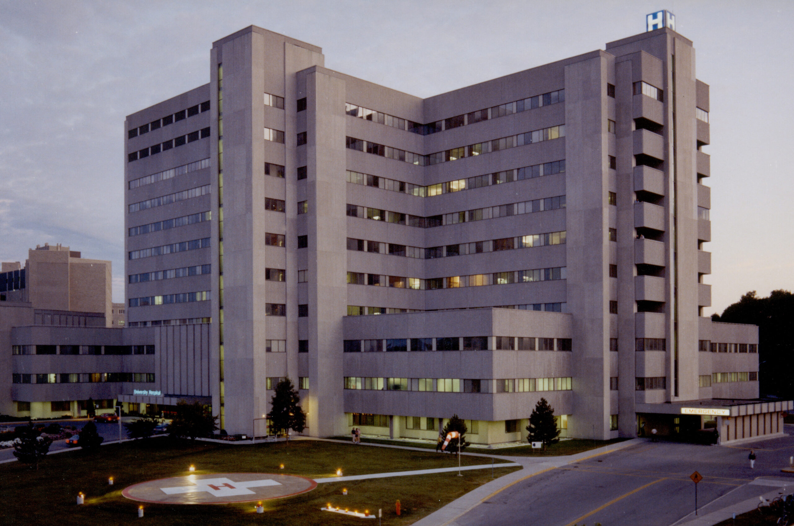
One area of care at London Health Sciences Centre (LHSC) that has a significant impact on the diagnosis and treatment of patients is Medical Imaging, which is a way to take pictures of the inside of your body and includes X-ray, Nuclear Medicine, Interventional Radiology, Ultrasound, Computed Tomography (CT) scan and Magnetic Resonance Imaging (MRI). This area of practice has changed dramatically since 1875 – from the early 1900s, when X-rays were first used on patients, to today, where the Medical Imaging department plays an essential role in a patient’s care journey. Medical imaging is used to diagnose conditions, guide interventions, and confirm treatment outcomes for patients across LHSC.
With over 300 staff who operate more than 100 imaging devices, performing over 450,000 imaging procedures each year for patients across our region, the team in Medical Imaging is frequently on the forefront of advances in technology helping transform patient care.
Improvements in technology have not just opened new areas or modalities for imaging and treatment, but have also enhanced imaging techniques, produced more detailed and precise images, and improved the ability to share them
Early 1900s – X-ray


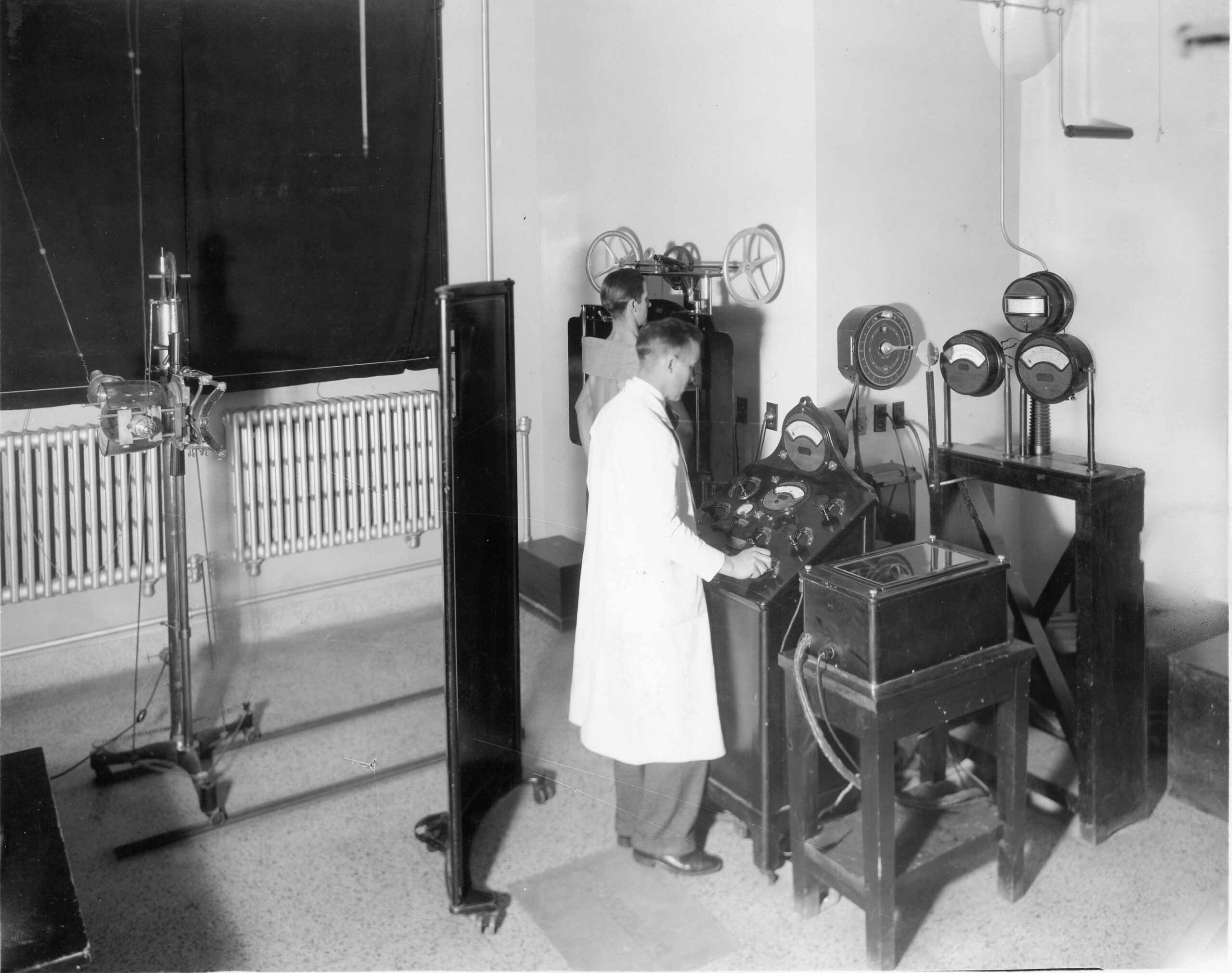
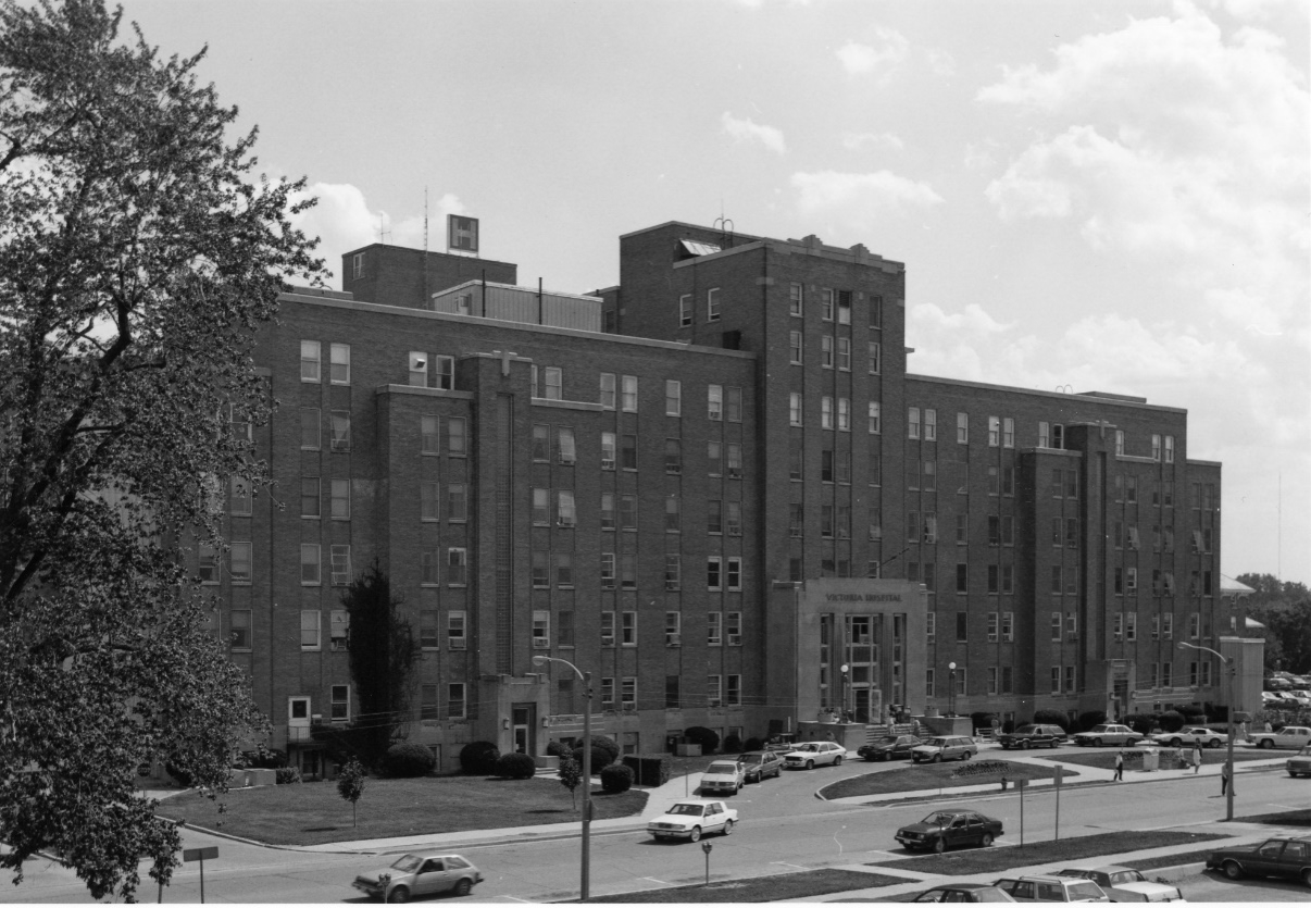
Above: Photo from 1930 of an X-ray being performed (left) compared to an X-ray room today in the paediatric Medical Imaging suite at Victoria Hospital with Jenn Andersen, a Medical Radiation Technologist (right).
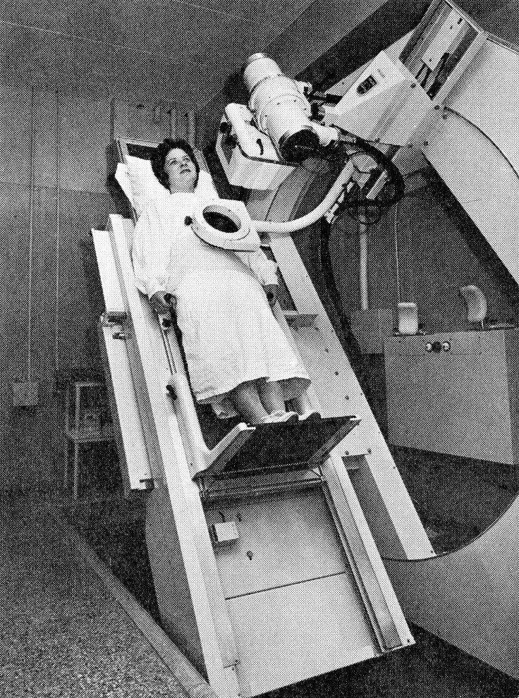
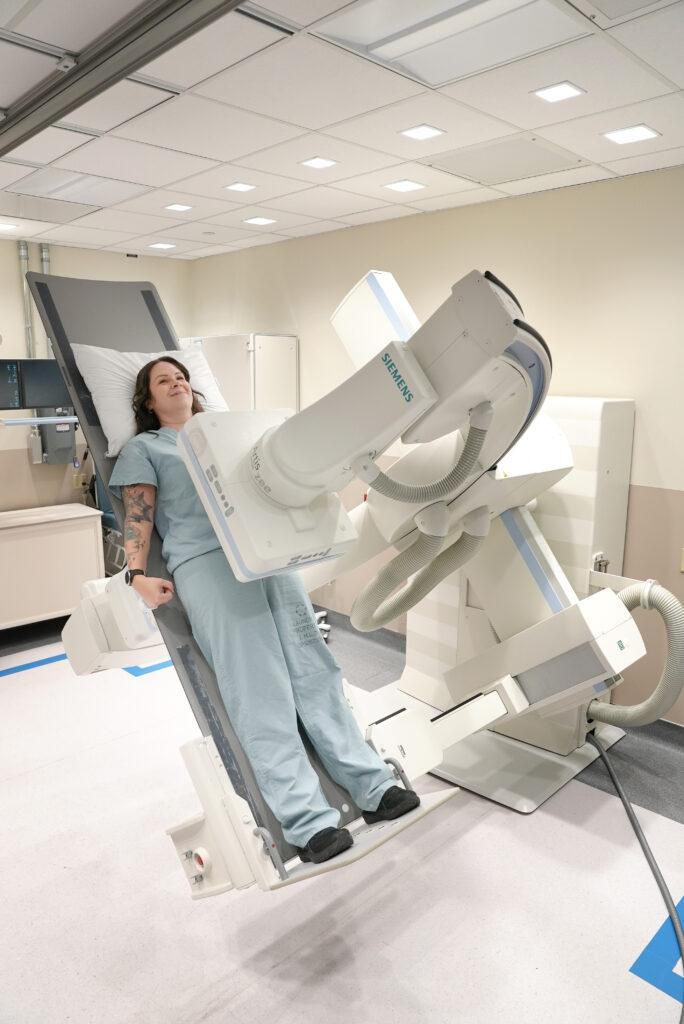
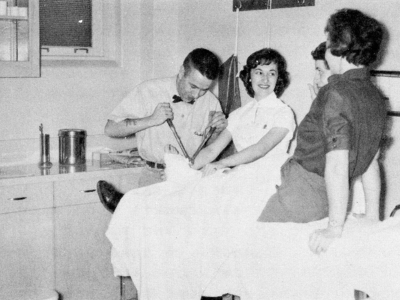
Above: A photo of fluoroscopy from 1962 (left) compared to the Fluoroscopy unit at Victoria Hospital today (right) with Melissa Morenz, a Medical Radiation Technologist, demonstrating how the procedure is performed.
- The first form of diagnostic imaging, X-ray has changed dramatically over the past 100 years. Today’s machines produce much lower doses of radiation with much clearer images.
- X-rays are a form of ionizing radiation used as a diagnosing tool. They are the most common tests done in Medical Imaging. An image is produced when a detector senses X-rays as they pass through the part of the body being imaged.
- Today, LHSC provides general radiology, Fluoroscopy and Bone Mineral Density scanning.
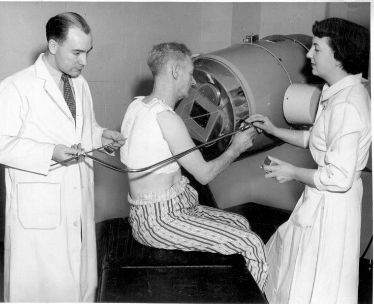
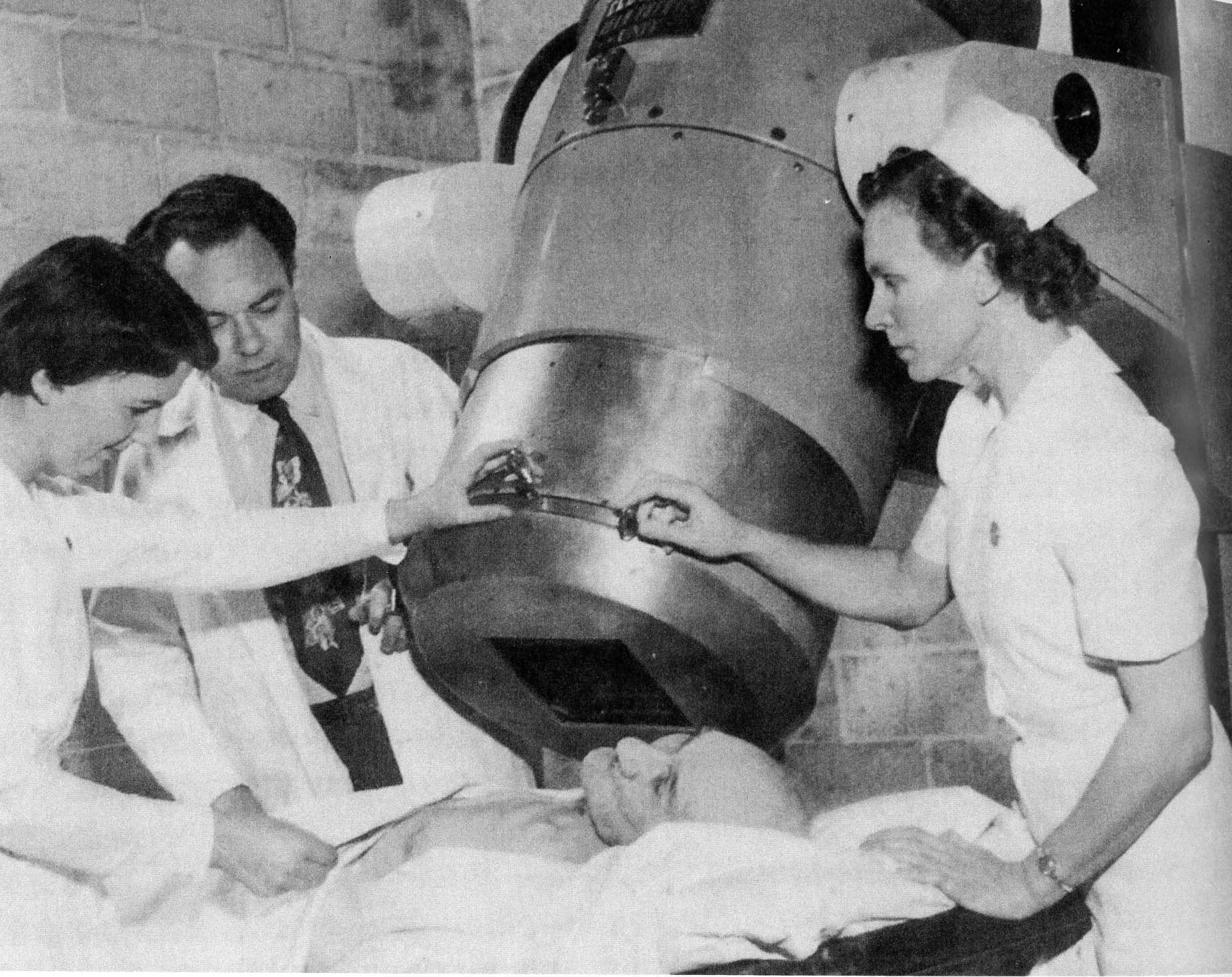
1950s – Nuclear Medicine (now Molecular Imaging and Theranostics)

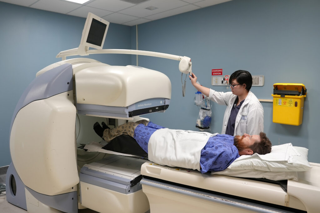
Above: A photo of a Medical Radiation Technologist adjusting the scanner in Nuclear Medicine in 1989 (left) compared to today’s Molecular Imaging and Theranostics’ suite (right) with Anne Vo, Medical Radiation Technologist, demonstrating the equipment.
- Nuclear Medicine began as a means of diagnosing and treating cancer at LHSC in the 1950s and has changed drastically over the past 25 years. Techniques now enable the targeting of specific cancer cells with radiation while leaving other cells alone.
- One of these advancements is the use of Positron Emission Tomography-Computed Tomography (PET-CT) scanning. In 2022, LHSC received its first PET-CT scanner at the Victoria Hospital campus. This scanner is used for diagnosis or staging of a wide variety of cancer types.
- Now known at LHSC as Molecular Imaging and Theranostics, this process uses small amounts of radioactive materials called radiotracers which are either injected into the bloodstream, inhaled or swallowed. The radiotracer travels to the area being examined and gives off energy in the form of gamma rays which are detected by a special camera and computer to create images to diagnose and treat disease. It treats the disease by attaching the radiotracer to a therapeutic isotope which releases a large amount of radiation to kill cancer cells, allowing for precise targeting and treatment of disease.
1970s – Interventional Radiology
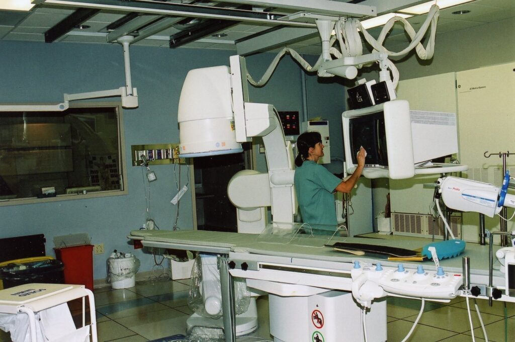
Above: An Interventional Radiology (IR) suite at University Hospital in the early 1990s (left) and the Neuro Radiology IR suite in 2025(right) with Elizabeth Cesarin, Medical Radiation Technologist.
- Interventional Radiology is a specialized field of medicine that uses advanced imaging technologies and devices to both diagnose and treat various medical conditions. By utilizing tools like X-rays, computed tomography (CT) scans, and ultrasound, Interventional Radiologists can precisely guide minimally invasive procedures.
- Interventional Radiology combines cutting-edge technology with medical expertise to perform minimally invasive procedures that often result in shorter recovery times compared to traditional surgery, using high-tech tools to navigate and fix problems inside the body.
- At University Hospital, the Interventional Radiology suite is dedicated to providing life-saving, minimally invasive care for patients with a wide range of urgent and complex conditions. This Neuro-Interventional Radiology suite is an essential component of LHSC’s designation as a Stroke Centre, while also supporting many other critical interventions.
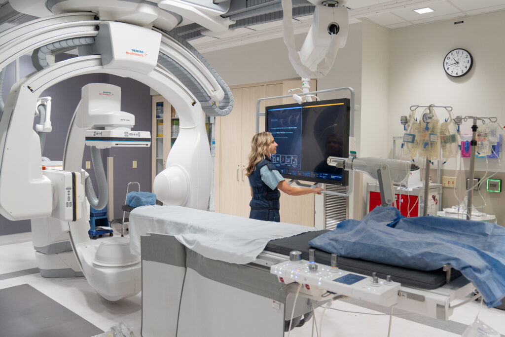
1970s – Ultrasound

Above: An image from 1983 with the opening of a new Ultrasound suite (left) and a recreation of this image today (right) with Leila Kawach, sonographer and Tracy Shao, student sonographer at Victoria Hospital.
- Ultrasound was introduced at Victoria Hospital in the late 1970s and later to University Hospital. Today, ultrasound is an essential tool that provides real time images, helping physicians and surgeons diagnose and treat many patients. Ultrasound imaging, also known as sonography, uses high frequency sound waves to create real time images of the inside of your body. It is commonly used to assess organs and vessels, monitor pregnancies, detect abnormalities, and assist with medical procedures.
- In 2023, LHSC took a major step forward in advanced imaging and obstetrical patient care with the acquisition of 12 new innovative ultrasound units.
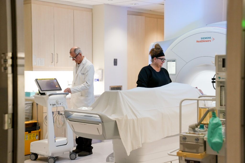
1970s/80s – Computed Tomography
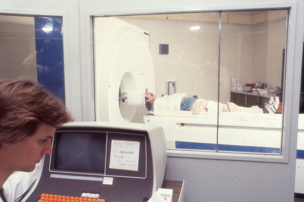
Above: A photo of CT scan in 1988 (left) and a recreation of the image today with Jason Lazar, Medical Radiation Technologist at University Hospital (right).
- One of the more recent modalities in medical imaging at LHSC is the CT Scanner. Installed in the late 1970s at University Hospital and at Victoria Hospital in the early 1980s, CT imaging uses a special X-ray system to produce images of a cross-section or slice of the body. The scanner captures images from multiple angles and the computer collects the information to make cross-sectional images. The two-dimensional (2D) images show a ‘slice’ of the anatomy. These individual slices can then be stacked and reconstructed to provide a more comprehensive evaluation of the area being examined.
- Currently, LHSC operates six CT scanners to support patients and research initiatives.
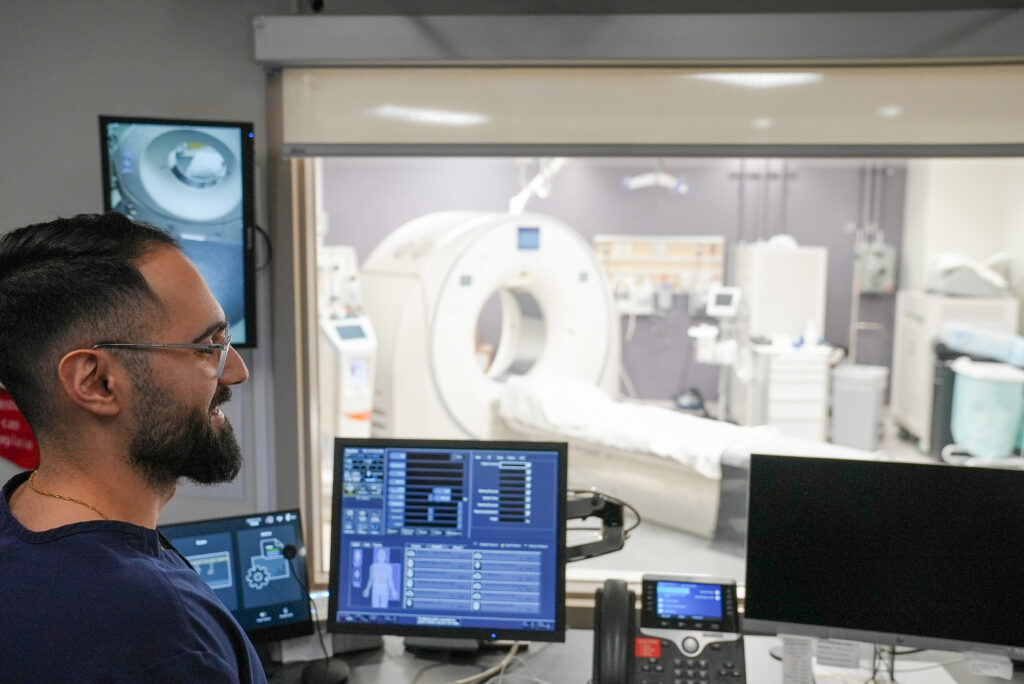
1980s – Magnetic Resonance Imaging

Above: An MRI is performed in 1986 at University Hospital with Dr. Donald Lee (left) and one is performed in 2025 with Dr. Lee and Michelle Smith, Medical Radiation Technologist (right).
- Magnetic Resonance Imaging (MRI) was introduced at LHSC in the 1980s. MRI uses a powerful magnet, radiofrequency waves, and computer technology to create cross-sectional images of internal organs and structures. Unlike X-Rays and CT scans, MRI does not use ionizing radiation.
- Over the years, LHSC has invested in upgrades to MRI technology. Today, LHSC has two 3 Tesla units, two 1.5 Tesla units, and one 0.55 Tesla unit.
LHSC is celebrating 150 years of care, innovation, and community impact by sharing 150 moments from our history. Join us in marking this milestone by sharing your own LHSC story.
Do you have an LHSC memory to share?
Submit your LHSC story today150 Moments
Celebrating the moments - big and small - that have defined LHSC's legacy
LHSC 150: Medical Imaging through the years
Honoring our legacy: A conversation with a former respiratory therapist
A new era of discovery: How 150 years of innovation has led to current research that is reshaping health care

Together, through every season: A holiday message
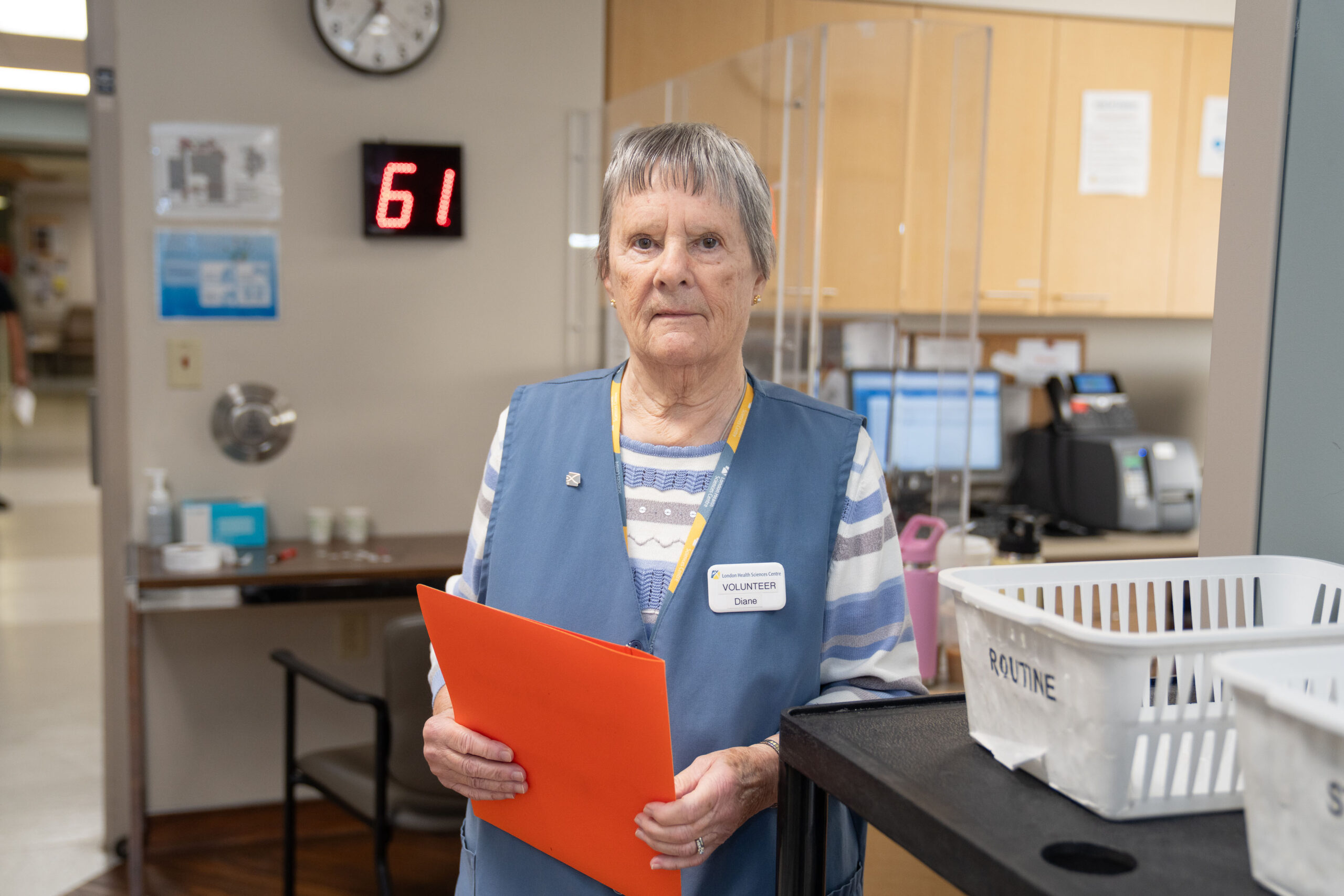
A lifelong commitment to volunteering: Celebrating 50 years of service
The evolution of Emergency Departments at LHSC
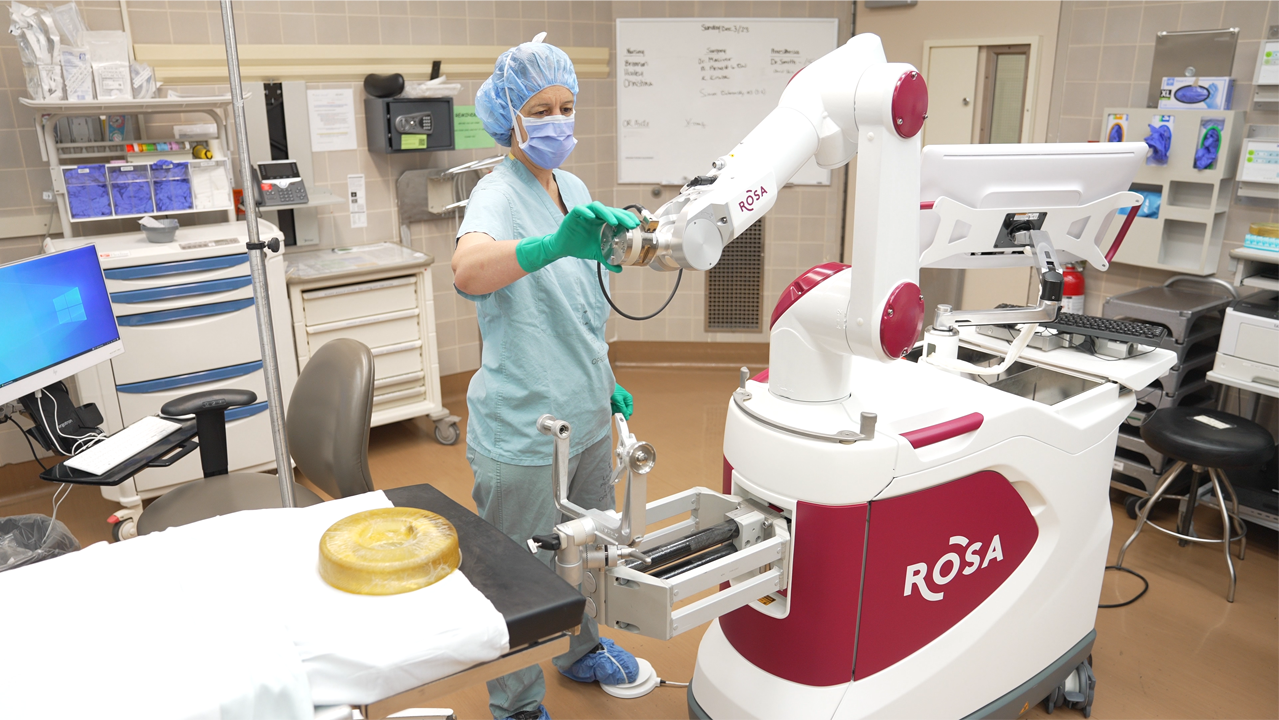
Meet your surgeon and their robot: The unstoppable duo behind cutting-edge surgeries at LHSC
LHSC 150: Patient Safety in leaps and bounds
Medical Device Reprocessing changes in sterilization procedures over the years
Helping kids be kids: A Children’s Hospital legacy
LHSC 150: Hockey and care through the years
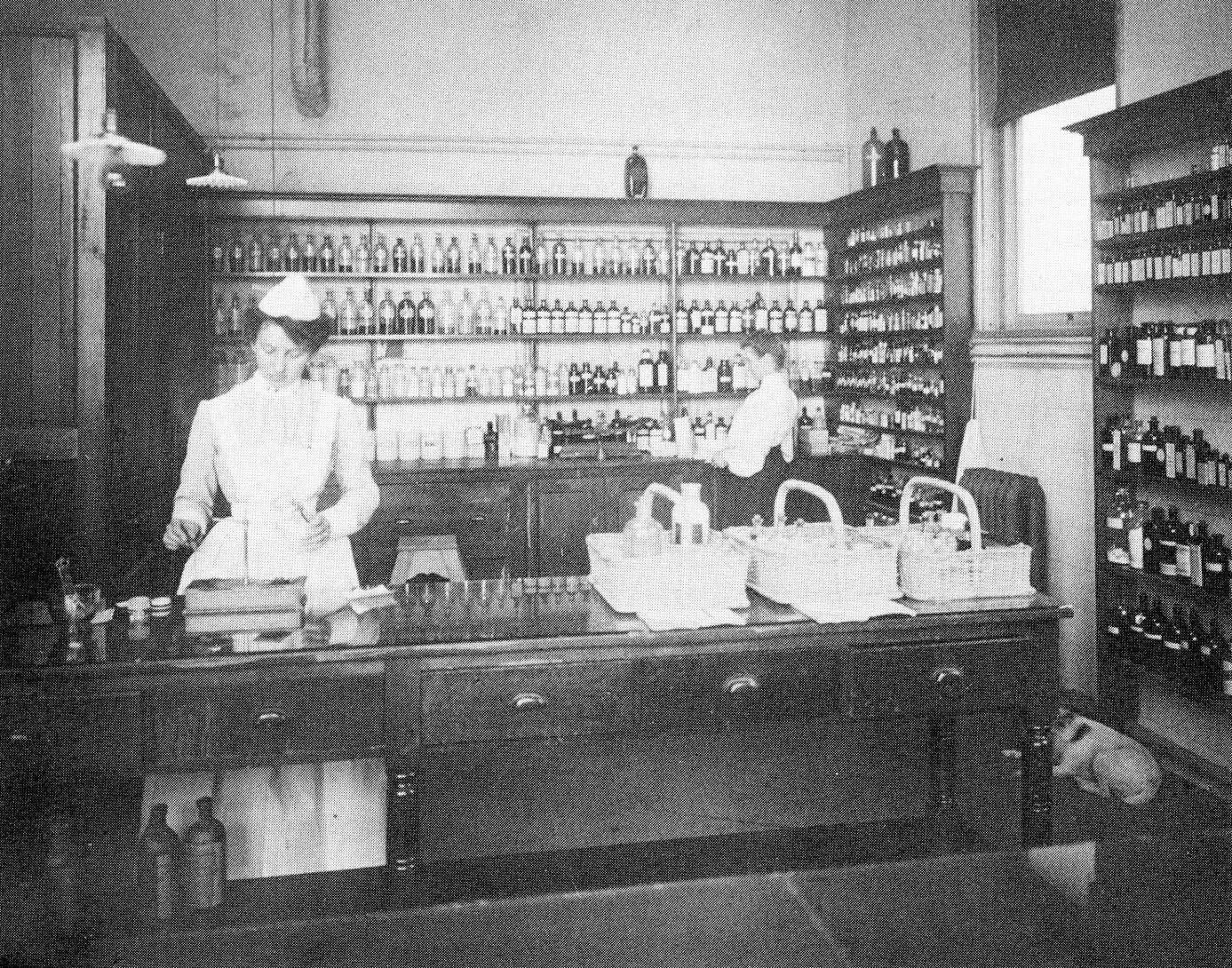
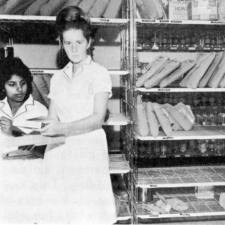
LHSC 150: The hospital pharmacy
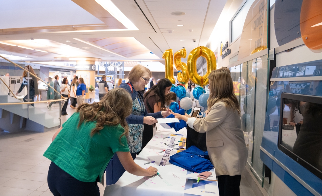
LHSC celebrates 150 years of great care, teaching, and research
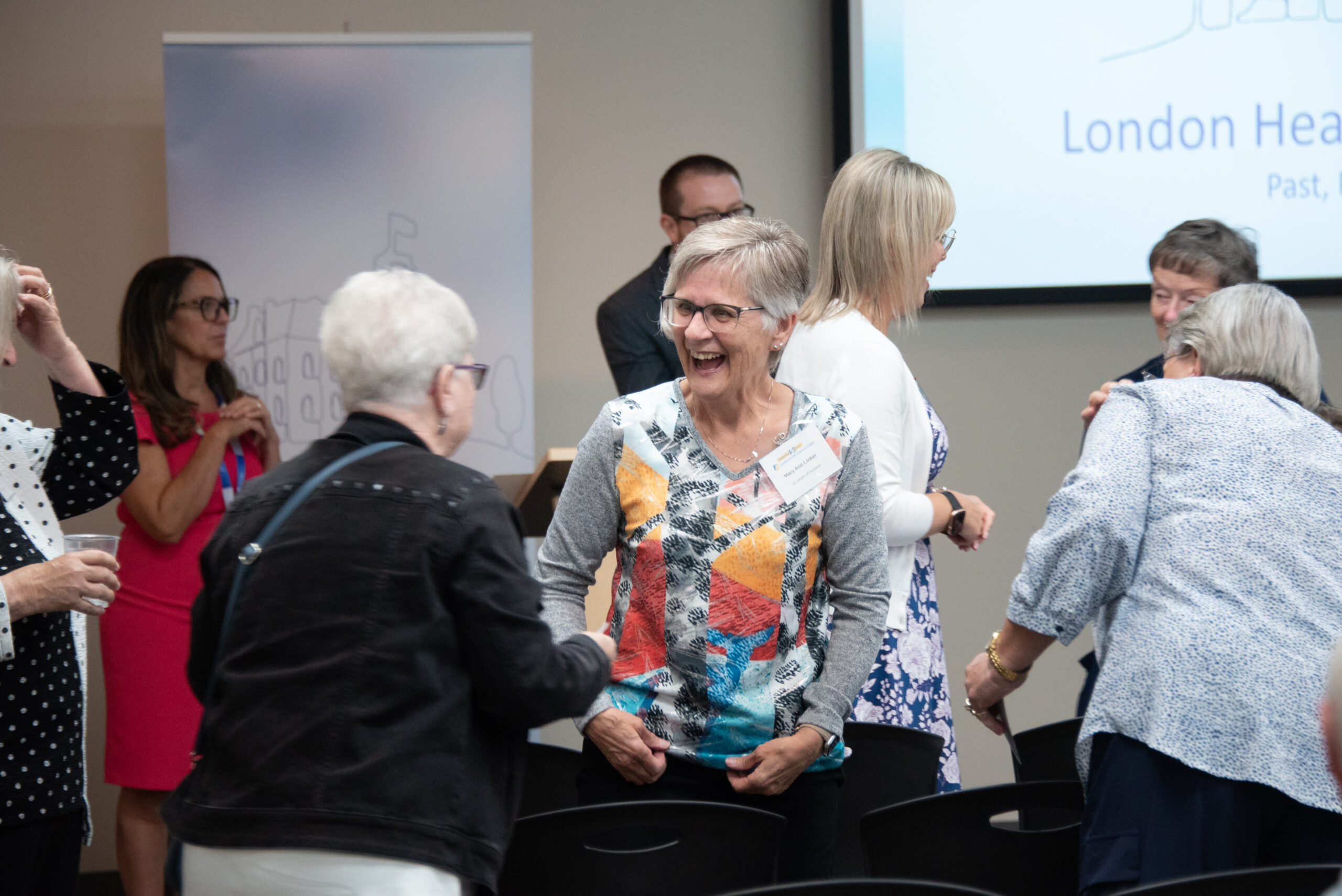
LHSC celebrates retirees who helped shape 150 years of great care, teaching, and research
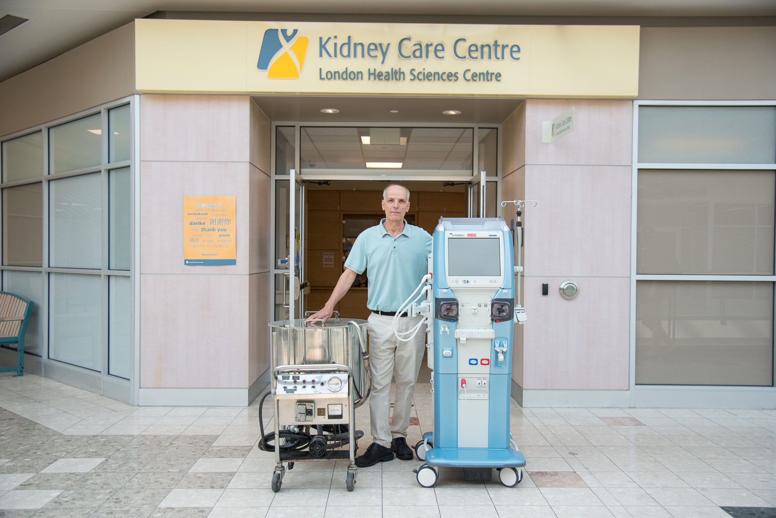
The first artificial kidney built in Canada has ties to London: A revolution in care for patients with kidney failure
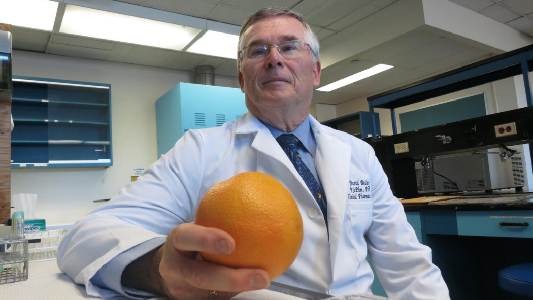
Citrus surprise: A juicy discovery at LHSC changed drug safety around the world
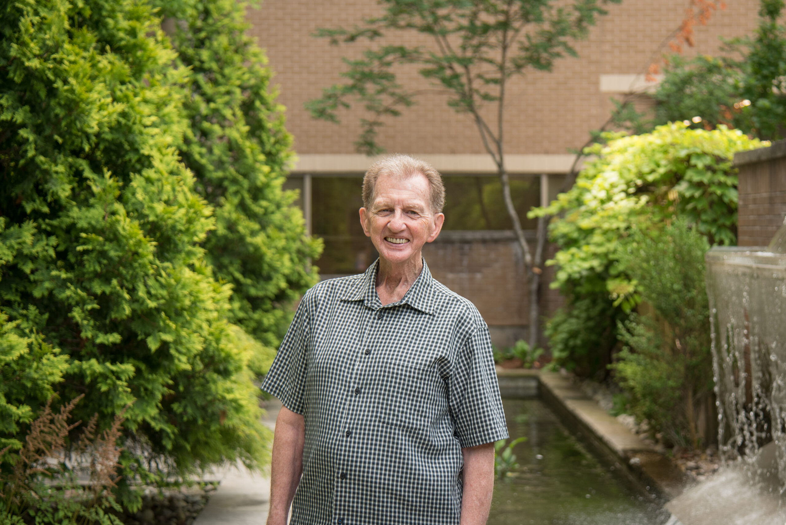
Gratitude and grace: Dave Gast’s 70-year health-care journey with LHSC
Join us for the LHSC 150 Anniversary Celebration
Join us for the LHSC 150 Retiree Tea
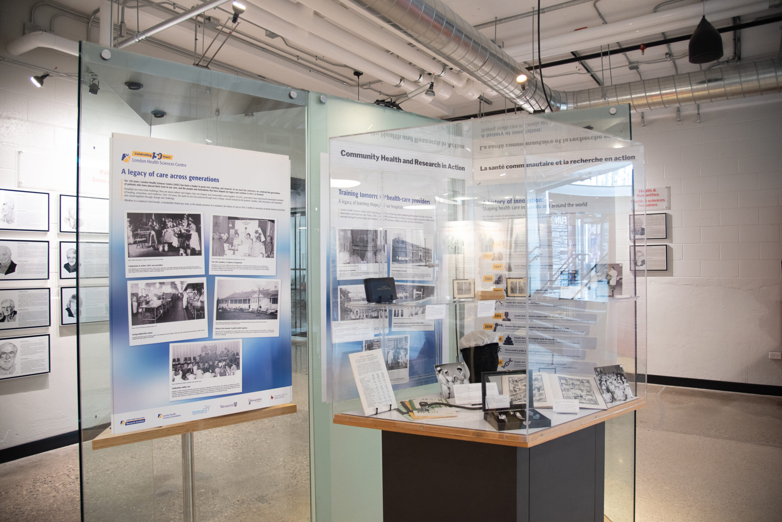
LHSC 150 Exhibit now open at the Canadian Medical Hall of Fame (100 Kellogg Lane)
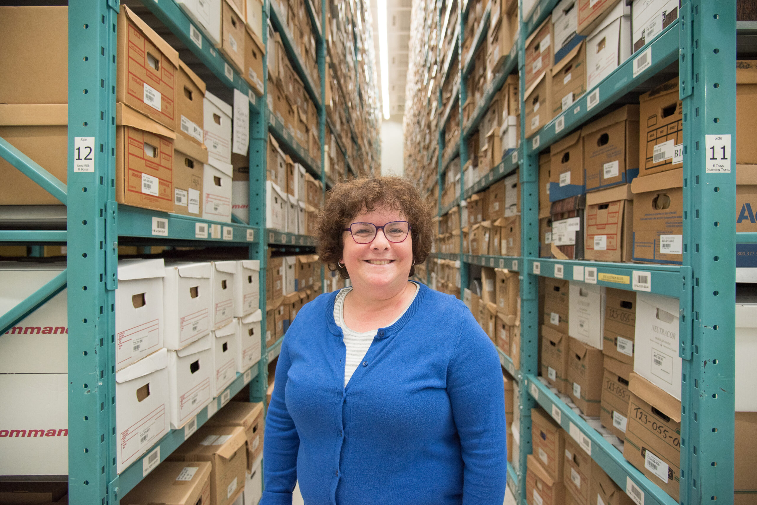
LHSC donates historic archives to Western Libraries
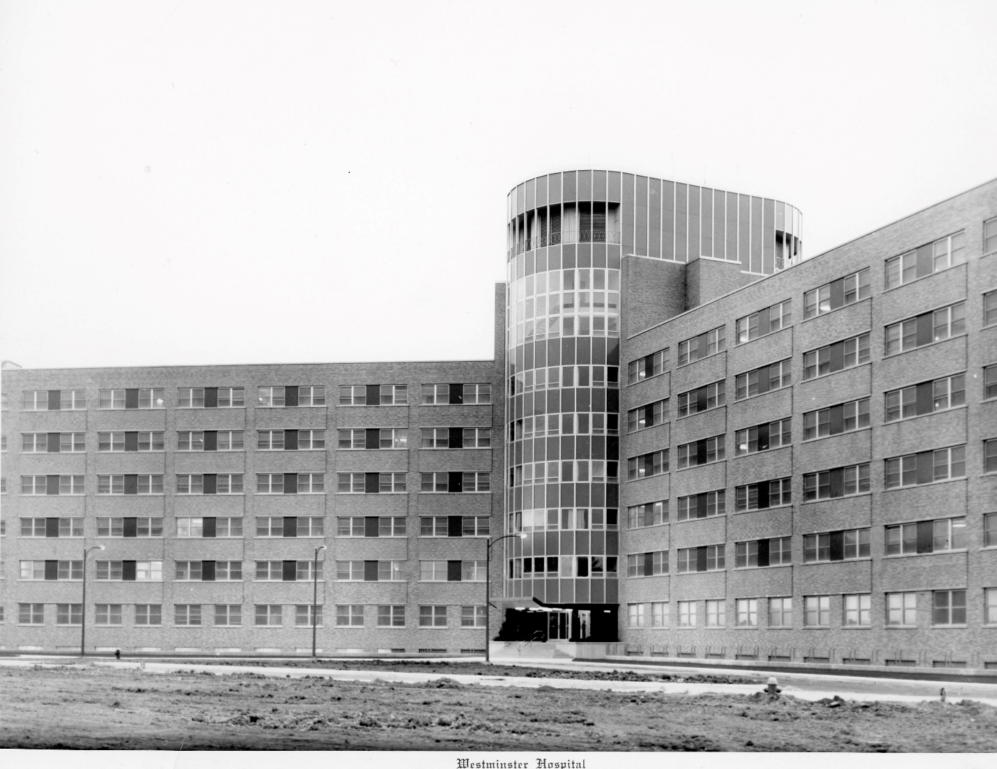
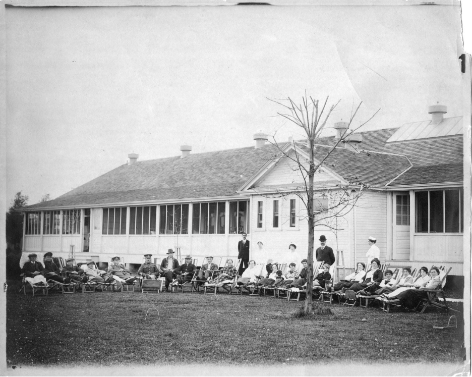
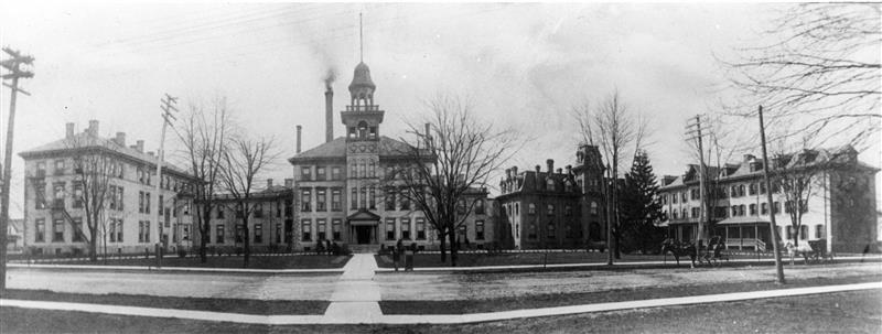
A new era of care: The journey from Westminster to Victoria Hospital
From impossible to lifesaving reality: Celebrating LHSC’s achievements in organ transplantation research
From the UH 50 Archives: Cardiac Care at heart of University Hospital
The history of cancer care at LHSC is a history of innovation
University Hospital: Ushering in a new era of care, teaching, and research
From the Cobalt Bomb to theranostics: LHSC’s pioneering role in cancer treatment
A history of support for children with physical, communication, and developmental needs
War Memorial Children’s Hospital: The history and legacy of paediatric care at LHSC
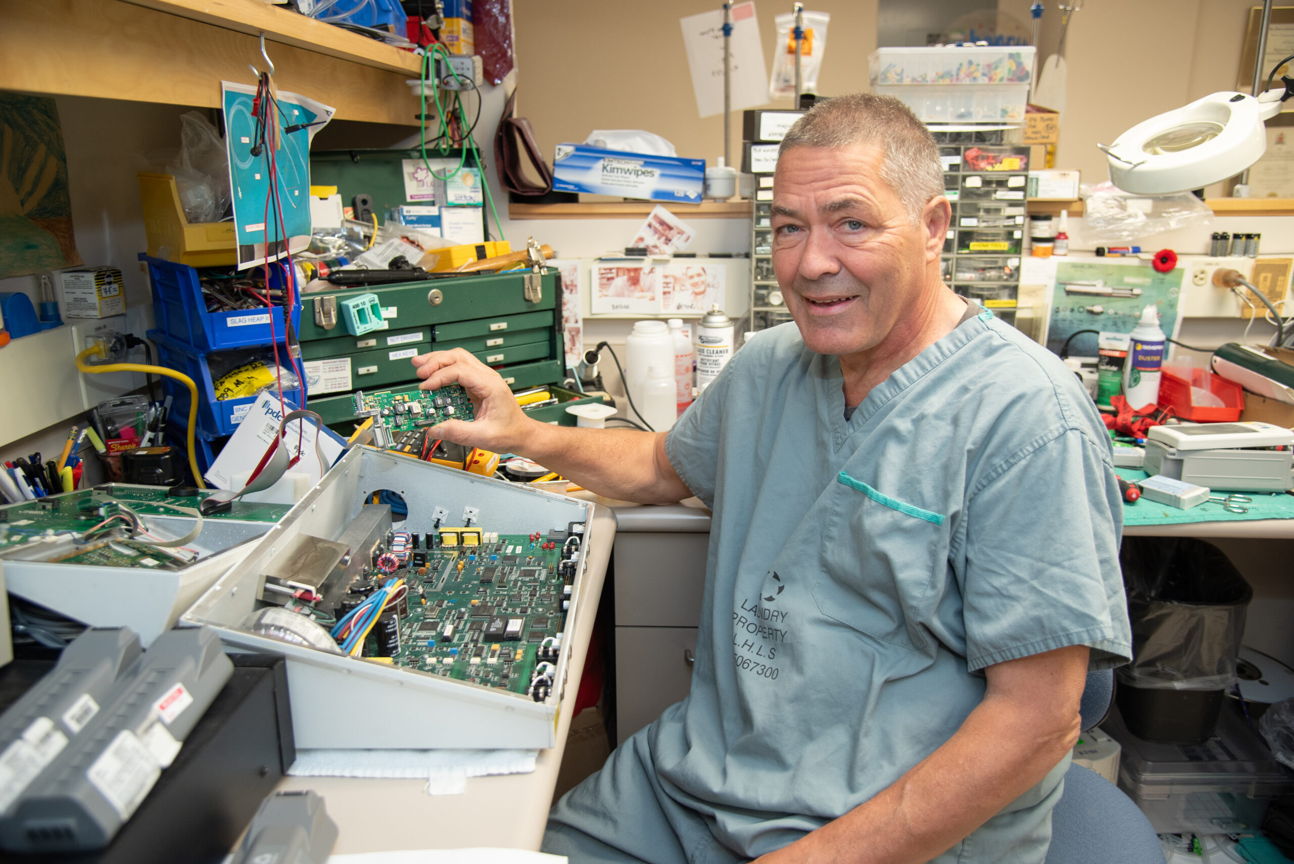
Advances in biomedical engineering

Celebrating 150 years of care during Nursing Week
Before LHSC: The community vision that transformed London’s health care
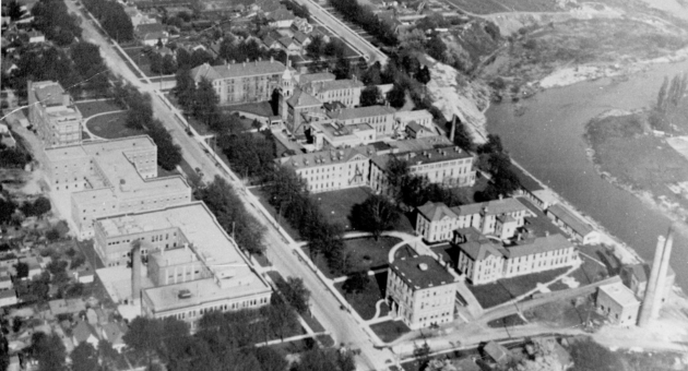
Victoria Hospital: A name that has shaped more than a century of care
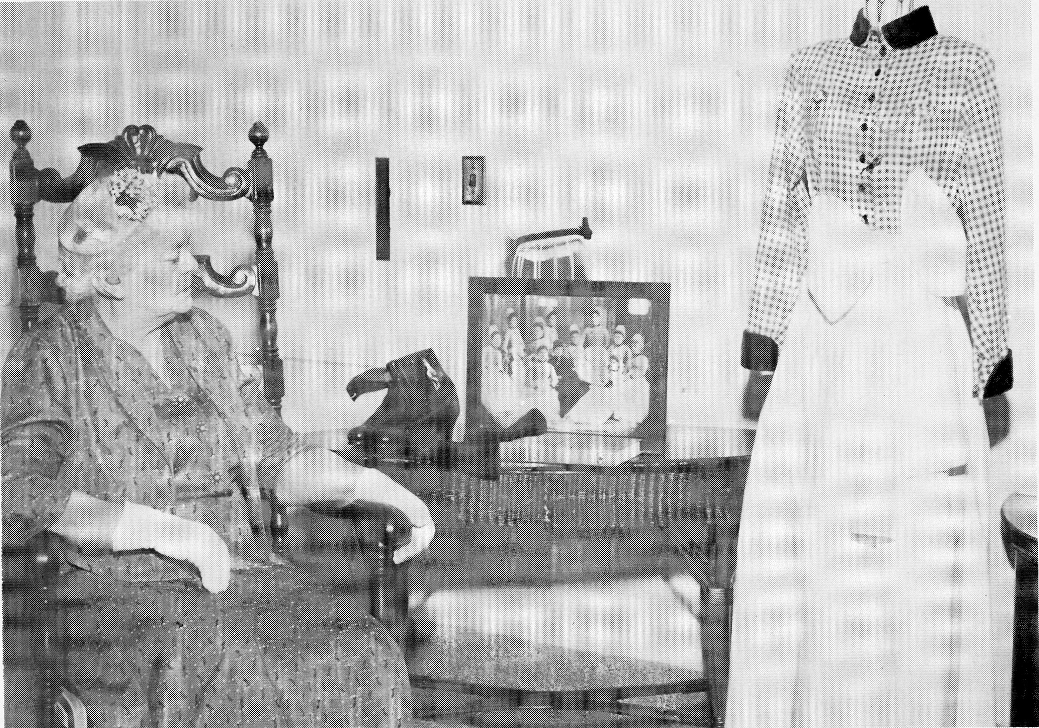
150 years of nursing education at LHSC: From the training school to education today
A legacy of innovation
Where it all began: The opening of London General Hospital
LHSC 150: Celebrating 150 years of great care, teaching, and research
%20--%3e%3cpath%20d='M196.8,535.6h806.4c35.6,0,64.4,28.8,64.4,64.4h0c0,35.6-28.8,64.4-64.4,64.4H196.8c-35.6,0-64.4-28.8-64.4-64.4h0c0-35.6,28.8-64.4,64.4-64.4Z'/%3e%3cpath%20d='M164.6,532.4l303.4-175.2c30.8-17.8,70.2-7.2,87.9,23.6h0c17.8,30.8,7.2,70.2-23.6,87.9l-303.4,175.2c-30.8,17.8-70.2,7.2-87.9-23.6h0c-17.8-30.8-7.2-70.2,23.6-87.9Z'/%3e%3cpath%20d='M468,830.9l-303.4-175.2c-30.8-17.8-41.3-57.2-23.6-87.9h0c17.8-30.8,57.1-41.3,87.9-23.6l303.4,175.2c30.8,17.8,41.3,57.2,23.6,87.9h0c-17.8,30.8-57.1,41.3-87.9,23.6Z'/%3e%3c/svg%3e)
%20--%3e%3cpath%20d='M1003.2,664.4H196.8c-35.6,0-64.4-28.8-64.4-64.4h0c0-35.6,28.8-64.4,64.4-64.4h806.4c35.6,0,64.4,28.8,64.4,64.4h0c0,35.6-28.8,64.4-64.4,64.4Z'/%3e%3cpath%20d='M971,643.9l-303.4-175.2c-30.8-17.8-41.3-57.2-23.6-87.9h0c17.8-30.8,57.1-41.3,87.9-23.6l303.4,175.2c30.8,17.8,41.3,57.2,23.6,87.9h0c-17.8,30.8-57.1,41.3-87.9,23.6Z'/%3e%3cpath%20d='M667.6,719.4l303.4-175.2c30.8-17.8,70.2-7.2,87.9,23.6h0c17.8,30.8,7.2,70.2-23.6,87.9l-303.4,175.2c-30.8,17.8-70.2,7.2-87.9-23.6h0c-17.8-30.8-7.2-70.2,23.6-87.9Z'/%3e%3c/svg%3e)