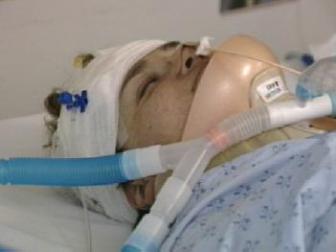
| Neurological Assessment | EEG and Continuous EEG |
| ICP monitor | Lumbar puncture (LP, Spinal Tap) |
| Nuclear Medicine | Peripheral Nerve Stimulator |
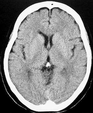
| CT Scan | Cerebral angiogram |
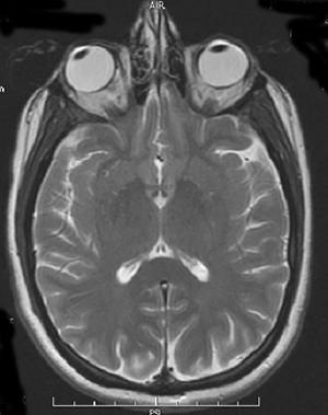
| MRI |
| Neurological Assessment | A doctor or a nurse can do a neurological assessment for a patient. The function of the brain, spine, and nerves is usually tested every twelve hours or more often if there are concerns. These tests can help keep track of changes in the health of the brain and nerves. |
|
An intracranial pressure (ICP) monitor is inserted by a neurosurgeon to monitor the pressure of the fluid (CSF) in and around the brain. It can also be used as a drain to remove extra CSF or blood and reduce the pressure on the brain. |
| Nuclear Medicine | Nuclear medicine involves using mildly radioactive substances, called isotopes, to identify injury or disease. It can be used to identify changes in blood vessels anywhere in the body. Certain radioactive substances will find and attach themselves to particular cell types or proteins. The person is then scanned with a special camera. The isotopes ‘light up’, telling the physicians where the cell or protein they are looking for has collected. This can be very helpful to identify areas that have been damaged by a stroke or hemorrhage. It can also be used to check if the treatment being given to open up blocked vessels has worked. Patients go to Nuclear Medicine with a nurse if they need this scan. |
|
A CT or CAT Scan is a specialized kind of X-ray. A person is scanned and the pictures gathered are turned into a 3D image by the machine’s computer. CT scans are good at picking up collections of fluid. Sometimes a patient is injected with an x-ray dye that will show up under the CT scan. This is called a CT with contrast. It can help narrow down where there may be problems with the vessels such as bleeding or blockages. A nurse goes with a patient to the Radiology department for CT scans.
|
|
MRIs are used to get a very detailed picture of the body tissues and organs, including the brain and spinal cord. MRIs use a strong electromagnetic field to make two and three dimensional images of the area scanned. The use of a strong magnet means it is important to remove all metal before an MRI. The scanner is loud, so patients and staff are given ear plugs. Sometimes a patient will be given a substance to increase the contrast and sharpen the details of certain body parts, such as the blood vessels or spinal cord. Like a CT scan, an MRI is painless. Patients travel to radiology with a nurse to get an MRI. |
|
|
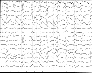
EEG is the short form for electroencephalogram. An EEG is a painless way to measure the electrical activity of the brain and may be used as a tool to diagnose brain function. There are a number of small stickers with a metal base called electrodes that are placed on specific areas of the head to monitor brain activity. An EEG is the only way to find seizures if there is no muscle activity. A person may be put on a continuous EEG if the team suspects the patient is having ‘silent seizures’. |
|
Lumbar puncture (LP, Spinal Tap) 
|
A lumbar puncture is a way to look for infection or blood in the brain and spinal cord. The patient lays on their side with their legs and arms curled up. The doctor gives them a local painkiller if needed and cleans the area with a solution to prevent infection.A needle is inserted between the lower back bones into the space next to the spinal cord. This space contains the same fluid (CSF) that slowly circulates around the brain and ventricles. A small amount of this fluid is drawn out and tested. Pressure is applied to the area to stop up any bleeding and a sterile dressing is applied. The results of tests done on CSF from a lumbar puncture can be used to decide what treatments may help. |
| Peripheral Nerve Stimulator | A peripheral nerve stimulator is used to make sure a patient is getting the lowest effective doses of certain medications that affect the nerves. The team wants to make sure they are sedated and pain-free using the minimal amount of drugs. This can help limit the possibility of side effects. |
|
|
Angiograms can be used to look at arteries anywhere in the body, such as the brain (cerebral angiogram) or heart (cardiac angiogram). Once the problem has been located, the physician may take steps to correct it all in one procedure. A cerebral angiogram can identify blockages that may have caused a stroke. They can also be used to find aneurysms. |