Procedure: Blood Gas Sampling
Ensure that patient and health care provider safety standards are met during this procedure including: - Risk assessment and appropriate PPE
- 4 Moments of Hand Hygiene
- Procedural Safety Pause is performed
- Two patient identification
- Safe patient handling practices
- Biomedical waste disposal policies
| Index - Refer to Procedure for Blood Sampling from an Indwelling Line
- Position Stopcock
- Assess SpO2
- Withdraw Blood Gas Sample
- Prevent Air Entry
- Label and Transport
- Flush the System
- Complete the Requisition
- Document
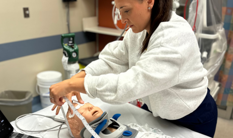
Equipment Required: 1 heparinized blood gas syringe 1 syringe or vacutainer for discard sample unsterile gauze square 1 chlorhexidine 2% and 70% alcohol swab PPE (non-sterile glove, mask with face shield) Functional arterial line | | PROCEDURE FOR BLOOD GAS SAMPLING | 1. | Following Procedure for Blood Withdrawal Access sampling port and draw a discard sample as per the procedure for Blood Sampling from an Indwelling Line. Review sampling considerations for arterial versus venous samples to avoid infusion contamination and document where sample was drawn | 2. | Position Stopcock Ensure stopcock is positioned to 45 degrees between syringe changes. Placement of the stopcock in the 45 degree position is "off" in all directions. This will prevent any flush solution from entering the tubing between syringe changes. | 3. | Assess Oxygenation and Ventilation Identify the patient's SpO2 reading at the time of blood sampling. If the patient is mechanically ventilated, record the minute volume (minute ventilation or Ve) from the ventilator. Record the minute volume when the patient is calm. The SaO2 result from the blood gas sample should be compared to the SpO2 that was obtained at the time the sample was drawn. The acid base balance is used to determine whether ventilation is adequate. A respiratory acidosis indicates that the total minute volume is not meeting the patient's needs, whereas, a respiratory alkalosis indicates that the minute volume is too high. To be meaningful, the gases should be compared to the minute volume that was obtained at the time the sample was drawn. | 4. | Withdraw Blood Gas Sample Slowly withdraw 1-2 mL into a heparinized blood gas syringe. Ensure that the syringe is securely attached to the access port. | 5. | Prevent Air Entry Hold the sample upright at a STRICT 90 degree angle with a gauze square over the top to prevent blood spatter. Gently tap any air bubbles to the top and expel into the gauze. Apply a cap to prevent blood contamination or air entry into the syringe. Do not draw air into the syringe to "level the air bubble" as you might with a medication. The PaO2 of room air is 140 mmHg; an air bubble will falsely increase the PaO2. | 6. | Label and Transport Label the syringe. Either take the sample to the point of care testing machine (GEM) or send the sample on ice to the lab in a biohazardous bag. Ice is only required for samples older than 30 minutes. | 7. | Flush the System Flush the catheter, stopcock and needleless access cap as per the Procedure for Blood Withdrawal. Replace the needleless access cap if blood cannot be completely cleared. Apply a new antiseptic sampling cap. Inadequate catheter flushing can lead to thromboembolism and loss of line patency. Thrombus formation is also a risk factor for vascular line infection. Residual blood in the stopcock or needleless access devices poses an infection risk. | 8. | Enter Ventilator Settings into Power Chart or GEM
Correctly identify whether the sample is arterial, venous or capillary. If venous, identify under comments if the sample is mixed venous, central venous or peripheral. Enter the patient's ventilator and temperature information into the lab requisition (for core lab samples) or the GEM program. Recording of ventilator settings enable correct interpretation of the patient's acid base balance and response to treatment. Blood gases are corrected to temperature. Include the following patient information for the core lab: - FiO2
- PEEP
- Ventilator mode
- PS/PC setting
- Minute Volume
- Patient Temperature
Include the following patient information for Point of Care testing in the core lab: - FiO2
- PEEP
- Minute Volume
- Patient Temperature
| 9. | Document Document results in the Electronic Health Record and communicate any significant findings to the provider and respiratory therapist. |
|
Developed: November 1988 (Morgan, B)
Last Reviewed: January 17, 2025 Brenda Morgan CNS, CCTC |