|
ECG; EKG; The heart produces an electrical signal. This signal can be picked up by placing small metal electrodes on the surface of the skin and measuring the electrical current that is flowing toward the electrode. This electrical picture of the heart's activity is called an Electrocardiogram. More frequently, we call this test an ECG, EKG or cardiogram. The electrical conduction through the heart follows a predictable and normal pattern (called Normal Sinus Rhythm). Normally, the current begins in the upper right chamber and travels downward toward the bottom left. This gives the ECG waveform a very specific pattern. Sometimes, different areas of the heart can initiate an electrical signal. If this happens, the electrical pattern will change. When the electrical current begins in a different area of the heart, the beat is called an "ectopic beat" or "arrhythmia". Most normal individuals will have occasional ectopic beats. We generally only treat ectopic beats or arrhythmias if the patient develops blood pressure problems. Patients in critical care have their ECG measured continuously. We usually will look at the rhythm from one or two different views of the heart. This allows us to identify arrhythmias and allows us to continuously monitor the number of times the heart beats per minute (heart rate). The ECG waveform and the heart rate is displayed on the bedside monitor (Image 1). We can also measure the flow of electricity through the heart from 12 (or more) different views or direction at a time. When we take all of these views, the test is called a "12-Lead ECG" (Image 2). When we look at the same conduction from different angles, we can identify important information about the function of the underlying heart muscle. For example, certain changes might tell us that the heart muscle is not getting enough oxygen, or that an area of the heart muscle has died (heart attack). Usually, the 12-lead ECG is a an intermittent measurement that is done with a portable machine. The 12-lead report is then stored in a central computer and reviewed by a cardiologist. We can also measure a 12-lead ECG continuously with the bedside monitor. |
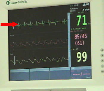
Image 1: Bedside ECG Monitoring |
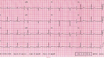
Image 2: A 12-Lead ECG |
|
|
|
|
|
|
Last Reviewed: October 23, 2014