|
MRI SCAN; MRA SCAN Magnetic Resonance Imaging (MRI) is a diagnostic test that produces very detailed pictures of body tissues and organs. MRI does not use xrays. Although CT is useful for certain types of images, MRI is better at detecting tissue details. The electromagnetic energy that is released when a patient is exposed to radiofrequency waves within a strong magnetic field is measured and analyzed by a computer to produce 2 and 3 dimensional images. MR angiography (also called MRA) is a special type of MRI study that specifically looks at the blood vessels. It is used to detect blood vessel disorders, providing detailed images without the use of standard contrast material. A special form of contrast may be used to make the images clearer. Like CT, the procedure is painless, however, conscious patients may experience claustrophobia or find the noise of the machine bothersome. Because the MRI will pull on any objects that contain metal, special precautions must be taken to remove anything with metal before the patient enters the MRI suite. This produces unique challenges when transporting critically ill patients who require invasive monitoring. It is important to let the nurse know if the patient has any implanted objects that contain metal. This includes pins or metal plates from orthopedic surgery, bullets or metal that is present from previous trauma or pacemakers. Patients must travel to the radiology department for an MRI/MRA. |
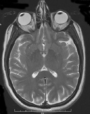
Image 1: Example of an MRI of the head. |
|
|
|
|
|
|
|
|
|
|
|