-
Introduction
-
Access Sites
-
Principles of CRRT
-
Artificial Kidney
-
Diffusion
-
Dialysis
-
Ultrafiltration
-
Hemofiltration
-
Pre/Post Dilution Hemofiltration
-
Clearance
-
Role of Filters in Clearance
-
Role of Diffusion in Clearance
-
Role of Hemofiltration in Clearance
-
Therapies Used
|
|
|
|
1.
|
INTRODUCTION
End-Stage Renal Disease (ESRD) refers to kidney disease that has resulted in the permanent destruction of a sufficient number of nephron units that renal function (waste and/or water removal) must replaced using an artificial kidney (Renal Replacement Therapy). Chronic dialysis is provided either using Intermittent Hemodialysis (IHD) or Peritoneal Dialysis. Chronic IHD is usually provided 2-4 days per week (depending upon the type of renal dysfunction). Peritoneal dialysis can be provided as Continuous Ambulatory Peritoneal Dialysis (CAPD) or Continuous Cycling Peritoneal Dialysis (CCPD). In CAPD, patients can administer and manage passive exchanges 3-5 times per day. CCPD requires patients to connect to a machine (usually at night) for automated exchanges.
Peritoneal dialysis is rarely used in critical care. Patients who are receiving peritoneal dialysis who are stable may have PD continued in the critical care unit. It will be run by a dialysis nurse who is trained in PD. Most critically ill patients who need Renal Replacement Therapy (CRRT) will receive either IHD or Continuous Renal Replacement Therapy (CRRT). Both intermittent hemodialysis and continuous hemodialysis circuits utilize the same principles. Blood is removed from the patient, pumped through a dialysis filter and returned to the patient following removal of surplus water and wastes. The filter performs many of the functions of the kidney's nephron unit, hence, it is referred to as an "artificial kidney".
The major difference between intermittent and continuous therapies is the speed at which water and wastes are removed. Intermittent hemodialysis removes large amounts of water and wastes in a short period of time (usually over 2-4 hours), whereas, continuous renal replacement therapies remove water and wastes at a slow rate more consistent of that of native renal function. While intermittent dialysis allows chronic renal failure patients to limit the amount of time that they are connected to a machine, the rapid clearance of solutes and fluid can be poorly tolerated when a patient is hemodynamically unstable.
During an acute illness, patients with ESRD often require more frequent renal replacement therapy to manage their increased production of metabolic by-products. Patients who develop Acute Kidney Injury that does not resolve with shock management may also require acute renal replacement therapy. RRT for either group may be provided using either IHD or CRRT. Hemodynamic stability usually determines the method.
Intermittent hemodialysis and SLEDD are both delivered using a conventional hemodialysis machine that creates dialysis fluid (called dialysate) by adding electrolytes and salts to city water that has been dechlorinated and purified using reverse osmosis (RO). Dialysate fluid is not IV sterile, therefore, it cannot be delivered into the blood path. IHD and SLEDD require an IHD and an RO machine and are only run by IHD trained nurses at London Health Sciences Centre.
In CCTC, CRRT is provided using a Baxter PrisMaxTM or PrismaFlexTM machine. CRRT is delivered using sterile fluids, therefore, solutions can be delivered as either dialysis fluid or as replacement fluids into the blood path.
|
|
2.
|
ACCESS
Historically, early dialysis circuits required the removal of blood from an artery with return of the "cleaned" blood to a vein. In the 1960's - 1970's, surgically implanted external arterial-to-venous shunts (e.g., Scribner, or Thomas shunts) were used for acute dialysis access. Early CRRT circuits required arterial and venous access devices (called Continuous Arterial-Venous circuits), as the arterial-venous blood pressure gradient was used to drive blood flow through the circuit. These arterial-venous CRRT circuits were fraught with challenges. Fluid removal could not be regulated , with effluent flow determined by the patency of the filter, the arterial-venous blood pressure gradient and the distance between the filter and the effluent collection bag. Over removal with hypotension often occurred at the start of treatment, with filter clotting imiting the duration of a treatment. With the introduction of a blood pump into the CRRT circuit, arterial-venous pressure gradients were no longer required for flow rates. This allowed temporary double lumen hemodialysis catheters to be introduced, eliminating the need for the surgical placement of arterial-to-venous shunts. This simplified initiation and lead to the development of the sophisticated technically available today. Although temporary circuits are all venous-venous today, red and blue labeling of catheter limbs and circuit tubing is universally used to identify the access limb (red is the usual side to remove blood) and return limb (blue denoting lumen where blood is returned).
Perm caths are double lumen dialysis catheters that are tunneled under the skin. They are made from material that is more appropriate for prolonged usage. Tunneling decreases the risk for infection or accidental dislodgement. The tunnelled portion of the catheter is separated from the external portion of the catheter by a dacron cuff which reduces migration of pathogens into the catheter tract and helps secure the device. Perm caths are usually inserted in Interventional Radiology Department. They are indicated for both prolonged dialysis in Acute Kidney Injury and ESRD.
ARTERIAL-VENOUS FISTULAS AND GRAFTS
Long term access for patients with ESRD may be created surgically through the use of an arterial-venous fistula or arterial-venous graft. Most are inserted in the upper limb, although, the lower limb can also be used.
An ARTERIAL-VENOUS FISTULA is the optimum access, created by suturing an artery and vein together. The capillary is bypassed, creating a high flow rate into the vein. This new circuit causes the vein to distend and bulge. It takes an average of 4 months before a fistula matures sufficiently to the point where it can be used for dialysis access. Consequently, for acute use, a temporary dialysis catheter may be required as a bridge until the fistula matures. At maturity, the fistula will change from being soft and pliable to firm and springy.
A buttonhole technique is typically used to access a fistula. Dull needles inserted repeatedly through the same hole will eventually cause a tunnelled tract, similar to an earing hole. Pain is minimized by using the same trac each time. The muscle layers of the artery close the tunnel after needle removal. Tunnel sites must be cleansed thoroughly and any scabs removed to prevent infection.
Fistulas are made from the patient's own biological material and are capable of functioning for many years. They are completely under the skin, which reduces infection when compared to double lumen catheters, and allows patients to swim and submerge in water. The high flow rates also reduce clotting
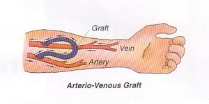
An ARTERIAL-VENOUS GRAFT is another surgical option, used when the vessels are too small to support the formation of a fistula. Graft material is used to create a conduit between the artery and vein. A graft requires less time to mature than a fistula, and may be accessed as early as 4 weeks after insertion. It is also easier to create. A graft is also associated with a lower rate of infection than a central venous catheter and allows the patient to swim.
Compared to fistulas, grafts do not survive as long but they are easier to implant. They also require needle access each time (fistulas do not generate tunneled tracks). Grafts are also more likely to clot.
Complications of fistulas or grafts include:
Infection
Clotting
Aneurysm (risk increases if the same tunnel cannot be used each time or if sharp needles are required)
Stenosis (arterial of venous)
Steal phenomena" (arterial flow being "stollen" from one area to another to "feed" the shunt).
ASSESSMENT AND MONITORING
Once mature, both fistulas and grafts should maintain a high rate of blood flow. This can be assessed by the following:
THRILL: A thrill is an "electrical sensation" or tingling created by the rapid flow of blood through the circuit. It can be felt by gently touching over top of the fistula.
BRUIT: A bruit is an audible whooshing sound that is heard when a stethoscope is placed gently over the fistula or shunt. This is created by the rapid flow of blood.
MONITOR: Watch for signs of complications including swelling at the site or distal extremity, redness or discharge.
AVOID: Avoid taking blood pressure, applying a tourniquet or constrictive clothing or excessive use of the affected limb.
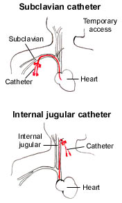
In critical care, temporary double-lumen venous dialysis catheters are the most common form of access. They can be inserted quickly at the bedside and used immediately. "Perm" catheters are double lumen venous catheters that are designed for longer indwelling use. They are used more frequently in patients with chronic renal failure and may be used as a bridge until a surgically created fistula is ready for use.
Dialysis catheters are easy to differentiate from regular intravenous lines by their red and blue hubs. The red lumen denotes the side of the venous catheter that is used to pull blood from the patient, and is referred to as the access lumen. The blue lumen is the return site and is used to reinfuse the patient's blood after it passes through the dialysis filter. If an adequate flow rate cannot be achieved by removing blood from the access side of a catheter, the catheter limbs can be reversed during dialysis. Reversal of the limbs does produce a small reduction in clearance due to recirculation that is not usually clinically important.
When a double lumen catheter is not in use for dialysis, some form of anticoagulant is always instilled into each lumen to maintain patency. While citrate is the most common agent, some catheters may still require blocking with heparin or tPA (particularly among patients with ESRD). If heparin is used, the concentration may be as high as 5,000 - 10,000 units per mL. Because each lumen contains a volume of ~1 to 2 ml, the two lumens could contain a maximum of up to 40,000 units of heparin! ALWAYS assume that each lumen contains full strength heparin (even if it is labeled as containing saline).
In CCTC, 4% Citrate is now the standard catheter blocking solution for dialysis catheters. Citrate binds to calcium to prevent clotting and does not affect the aPTT. Citrate is the standard for blocking all CRRT catheters in CCTC, even when heparin has been used to maintain filter patency.
EMERGENCY VASCULAR ACCESS:
A double-lumen venous dialysis catheter can be used as a central venous infusion site during an emergency. To ensure the line remains patent for subsequent dialysis treatments and to reduce the risk for infection, these lines should only be used for dialysis.
If a dialysis catheter is the only vascular access available in a life-threatening emergency, it can be used as a central line. HOWEVER, always assume that the catheter contains heparin, TPA or another agent. Aspirate at least 5 ml from each lumen and flush vigorously before connecting to an infusion pump to maintain patency. Maintain sterility to preserve the site for future access.
|
|
3.
|
PRINCIPLES
Hemodialysis employs the principles of diffusion, hemofiltration and convection, using an external filter to create an artificial nephron unit.
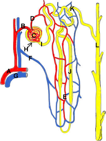
Recall the normal nephron unit:
|
|
Blood flows from the Renal Artery (A) to the Afferent Arteriole (B). The Afferent Arteriole then enters the Bowman's Capsule (H) and becomes the Glomerulus (C). Blood leaves the Glomerulus via the Efferent Arteriole (D), which continues to become the Peritubular Capillary (E).
Water and solutes that are filtered through the Glomerular Membrane collect in the Bowman's Capsule (H) and drain into the Proximal Tubule (I). Filtrate continues through the Loop of Henle (J), Distal Tubule (K) and Collecting Tubule (L). Water and solutes are reabsorbed from the filtrate into the peritubular capillaries, while solutes can also be secreted from the peritubular blood into the tubule system for elimination in the final urine.
|
The diagram above depicts one nephron unit. Each kidney has approximately one million of these microscopic units. They collectively maintain water, electrolyte, waste and acid-base balance.
Arterial blood flows from the renal artery, branching into smaller divisions known as arterioles. Branches of the arterioles eventually carry blood into small "containers" called Bowman's Capsules, located in the cortex of the kidney. The arteriole that Arrives at the Bowman's Capsule is called the Afferent arteriole. The blood then flows into a specialized capillary (located inside the Bowman's Capsule), called the GLOMERULUS. Any blood remaining at the end of the glomerulus Exits the Bowman's Capsule via the Efferent arteriole.
The afferent arteriole is larger in diameter than the narrow efferent arteriole. This arrangement provides a high rate of blood flow into the glomerulus, but a high level of resistance to blood flowing out of the glomerulus. This structural difference produces a hydrostatic pressure within the glomerulus that is twice that of other capilliaries in the body. This increased hydrostatic pressure forces more water to move from the glomerulus, across the semi-permeable glomerular membrane and into the Bowman's Capsule.
Particle that are small enough to pass through the glomerular membrane will diffuse from an area of high concentration (from the glomerulus) to low concentration (to the Bowman's Capsule). When large volumes of water are forced across the membrane, additional particles (or solutes) are "dragged along with the water". Thus, the large movement of water across the glomerulus removes even more solutes than diffusion alone would remove. The "washing" of additional solutes across the membrane by a large flux of water is known as convection.
Proteins are larger molecules and are too big to fit across a normal glomerular membrane. Consequently, blood that leaves the glomerulus via the efferent arteriole has most of the water and electrolytes removed, but all of the plasma proteins remaining. Thus, blood in the efferent arteriole has a higher oncotic pressure.
In order to adequately eliminate all of the waste products produced each day, we have to filter very large volumes of water across the glomerulus. About 1200 ml per minute of filtrate is produced. By the time enough water has been moved across the membrane to wash out all of the surplus waste products, over-removal of water, glucose, electrolytes and other substances has occurred. Consequently, large amounts of the filtered water and solutes will need to be reabsorbed from the tubule fluid into the blood. Solutes and water are reabsorbed into capillaries that are wrapped around the tubules, called peritubular capillaries. These peritubular capillaries are the continuation of the efferent arterioles. They are also responsible for perfusing the kidney. In addition to reabsorbing water and solutes from the tubule filtrate, surplus solutes can be secreted from the peritubular capillaries into the tubule filtrate for elimination in the urine.
|
|
4.
|
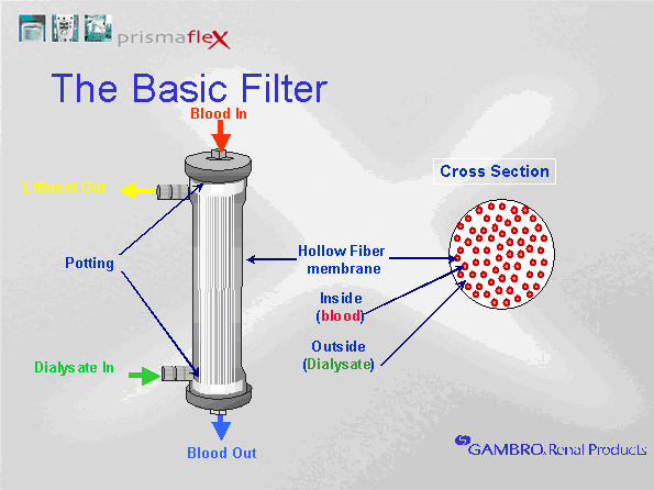
ARTIFICIAL KIDNEY
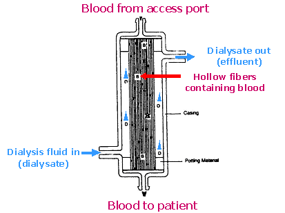
The dialysis filter is referred to as an artificial kidney. Blood is pulled from the patient and carried into the filter. Once inside, the blood travels through many tiny tubules called hollow fibers. Water and solutes can pass across the semi-permeable membrane between the blood and the fluid that surrounds the hollow fibers. Any fluid or solutes that enters the filter canister will be drained out as waste.
Schematic of dialysis filter (artificial kidney)
Note how the dialysis filter has structural similarities to the nephron unit. Blood arrives at the filter via the access tubing (afferent arteriole). Blood enters the small hollow fibers within the filter (glomerulus). Water and solutes diffuse across the semi-permeable membrane of the hollow fibers and collect in the canister (Bowman's Capsule). Collected fluid (filtrate or effluent) is then removed via the drainage tubing (collecting tubule). Blood that remains in the hollow fibers is returned to the patient via the return side of the filter (efferent arterial).
Although similarities exist between the nephron unit and the artificial kidney, the artificial kidney has limited capabilities. In the nephron unit, filtered water and waste enters the proximal tubule. Because the nephron unit removes significantly more water and solutes than needed, most of the water and electrolytes that enter the tubule system are reabsorbed.
Unlike the nephron unit, the artificial kidney cannot reabsorb water or solutes that enter the filter canister Any filtrate that enters the filter canister will be removed via the drainage tubule. Consequently, one of the differences in the artificial kidney is the absence of the proximal tubule, Loop of Henle and distal tubule where water and solute reabsorption and secretion occurs. Thus, the drainage tubule that exits the filter is similar to the collecting tubule of the nephron unit, not the proximal tubule. To compensate for the inability to reabsorb water and solutes following removal from the blood, the artificial kidney is manipulated to restrict the actual removal to only surplus water and wastes. This is done by adjusting dialysis solutions and ultrafiltration rates. If more water or solutes are removed than desired, they may need to be given back via intravenous infusions.
The artificial kidney does not replace other important kidney functions, including stimulation of red blood cell production (erythropoietin), blood pressure and sodium regulation (renin) and calcium uptake by the GI tract (vitamin D synthesis). The nephron normally traps and recycles bicarbonate to maintain acid base balance. Bicarb is given to patients during hemodialysis to compensate for bicarb deficits.
The principles used during hemodialysis are reviewed below:
|
|
5.
|
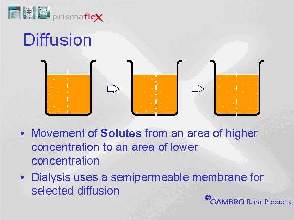
DIFFUSION
Diffusion is the movement of particles (solutes) across a semi-permeable membrane. Diffusion is the movement f
From the side with the highest concentration of particles, to the side with the lowest concentration.
|
|
6.
|
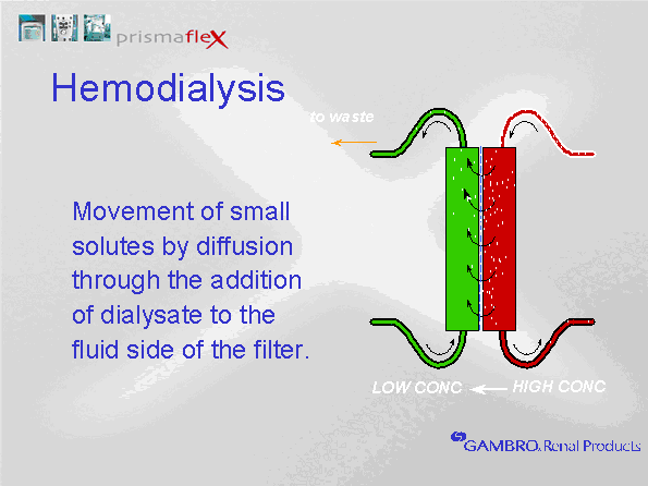
DIALYSIS FLUID (DIALYSATE):
Dialysate is the fluid that is pumped into the filter canister, surrounding the hollow fibers. The concentration of solutes in the dialysis fluid determines diffusion gradients. The removal of surplus solutes from the blood is achieved by infusing dialysate fluid that contains a lower solute concentration than the serum concentration (e.g. dialysate does not contain urea or creatinine).
To maintain normal serum electrolyte levels, dialysate fluid contains sodium, chloride and magnesium levels that are equal to serum concentrations (thus, removal of these electrolytes should only occur if the blood level exceeds normal serum concentrations). In renal failure, potassium is often high at the start of a treatment, therefore, we may begin dialysis with a low concentration of potassium in the dialysate. Because potassium is easily removed during dialysis, and continued dialysis will be required to ensure removal of other wastes such as urea and creatinine, potassium concentrations in the dialysate often require upward adjustment as the potassium level in the blood falls. Although in theory, potassium levels should not fall below 4 mmol/L in the serum if the dialysate contains 4 mmol/L, a number of factors influence serum potassium levels in critical care. Insulin therapy and the use of sympathomimetic drugs promotes the movement of potassium from the blood into the cells. This can lower serum levels. Additionally, potassium loss through the GI tract can increase the potential for hypokalemia. Low magnesium levels will also suppress the serum potassium levels, therefore, magnesium deficits should be replaced as needed. Additionally, high hemofiltration rates can lead to additional potassium clearance. Potassium levels must be monitored closely and adjusted to maintain normal serum concentrations.
In renal failure, serum bicarbonate levels are generally low, therefore, a source of bicarbonate is added to the dialysate to facilitate diffusion of bicarbonate into the blood. At one time, lactate based products were used for CRRT. Lactate is metabolized to bicarbonate to provide a less expensive source of bicarbonate replacement with longer product stability. Because critically ill patients with organ dysfuncition will often have impaired hepatic clearance, lactate based products were frequently associated with prolonged lactate elevation. We no longer use lactate-based products.
All dialysis solutions that are currently in use in CCTC are now bicarbonate based. We use 3 primary solutions, all contain 32 mmol/L of bicarbonate after mixing. The bicarbonate is contained in an upper compartment of a 5 L dialysis bag. To release the bicarbonate, squeeze the upper compartment until the two compartments mix. Failure to mix the two compartments can result in severe electrolyte abnormalities. The contents in compartment A, B and A plus B are listed on each bag. As well, failure to mix the two compartments would mean that only the volume in the lower compartment would be accessible. The scale would still weigh the bag and attempt to "draw" the upper compartment volume, pulling air into the circuit.
Dialysis fluid does not in theory cross into the blood side of the filter. It runs along the outside of the blood filter and into the effluent drainage. Whatever dialysis volume is administered, an equal volume will be removed in the effluent. This is automatic (dialysate input = dialysate output). Recall that hemodialysis solution is not IV sterile in IHD. For this reason, some fluid removal must always be maintained to prevent any crossing of dialysis fluid to the blood side. In CRRT, solutions are IV sterile. Therefore, patient fluid removal can be turned to zero because there is no risk of infection should dialysis fluid enter the blood path.
When patient fluid removal is set to zero, the hourly fluid removal might be slightly positive (usually 1-5 ml per hour). This actually indicates a gain of patient fluid by 1-5 ml.
Concentration gradients play a major role in diffusion. These will be explored further in the discussion on clearance. The other factor that influences diffusion is the type of filter used and the size of the molecule to be cleared. Diffusion of solutes cannot occur across a concentration gradient if the pore size is too small to permit passage.
|
|
7.
|
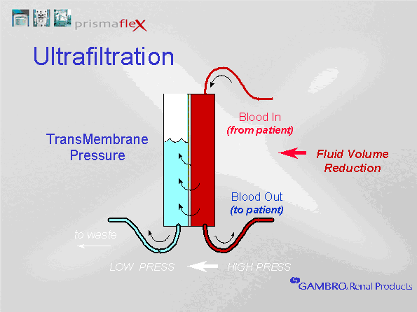
ULTRAFILTRATION
Ultrafiltration is the movement of water across a semi-permeable membrane because of a pressure gradient (hydrostatic, osmotic or oncotic). The increased blood pressure in the glomerulus creates a favourable driving pressure to force water across the glomerular membrane.
Blood pressure within the hollow fibers is positive, while the pressure outside the hollow fibers is lower. Increased negativity can be generated outside the hollow fibers by the effluent pump by either increasing the fluid removal rate, or by increasing the replacement flow rate. The difference between the blood pressure in the hollow fibers and the surrounding pressure is the Transmembrane Pressure (TMP). The TMP determines the ultrafiltrate production.
Different filter membrane properties can produce different ultrafiltration rates at a constant TMP. A filter that is more permeable to water will allow more water to travel across the membrane at a given TMP. A filter with a high permeability to water is called a high flux membrane.
|
|
8.
|
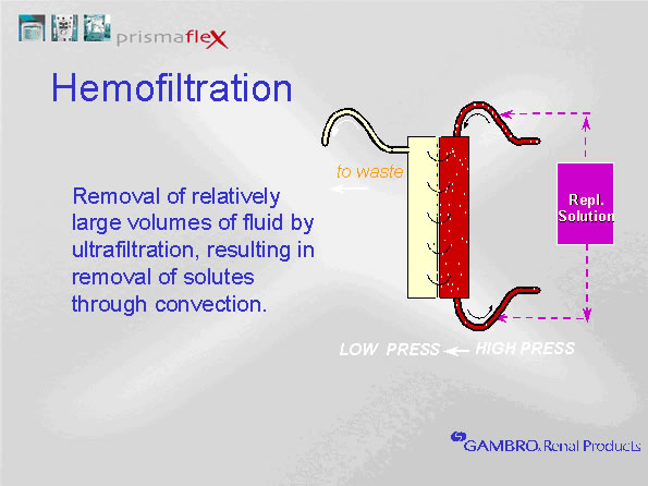
HEMOFILTRATION
In hemodialysis circuits, pulling large volumes of water across the semi-permeable membrane creates a convective current that "drags" additional solutes. While diffusion is effective at removing most small molecules, convection enhances the removal of small and mid-sized molecules. Thus, convection can be added to hemodialysis therapy to enhance solute removal. To prevent hypovolemia, any water removed during hemofiltration must be returned to the blood before it reaches the patient. This is called "replacement" fluid. Hemofiltration rates of 1 L/hr mean that one liter of fluid is removed from the patient's blood and eliminated in the drainage fluid AND 1 L of replacement fluid is returned to the circuit before it reaches the patient. We set hemofiltration rates by adjusting replacement rates. Any fluid removed during hemofiltration is given back to maintain a net neutral fluid balance. Replacement fluid must be sterile intravenous fluids with concentrations of electrolytes similar to plasma.
For example, if the CRRT therapy includes a hemofiltration rate of 1 L per hour, and the fluid removal is set at 200 ml per hour, 1200 ml will be pulled from the patient and introduced into the drainage collection bag each hour. Because the 1 L of hemofiltration is replaced, the net fluid removed is 200 ml. Whether hemofiltration is used or not, the net fluid removed is equal to the fluid removal setting.
|
|
9.
|
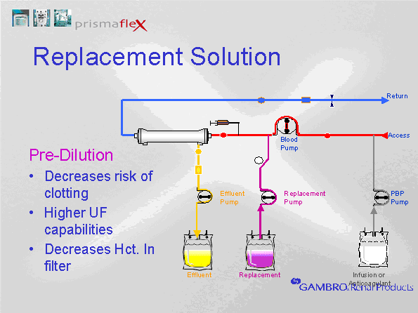
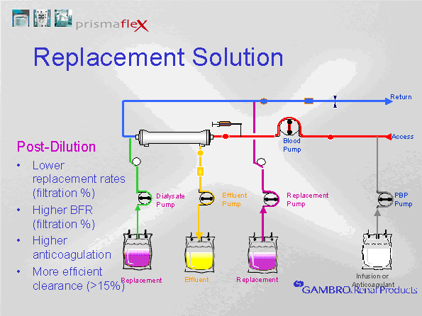
PREDILUTION VERSUS POSTDILUTION HEMOFILTRATION
Replacement fluids can be returned either pre or post filter. This is referred to as predilution or post dilution sets. Predilution means that the replacement solution is returned to the blood before it reaches the filter, diluting the blood in the hollow fibers. Postdilution means that the replacement fluid is returned to the blood after the filter (but before the return side of the access catheter). Predilution dilutes the blood in the filter, reducing clotting. Postdilution concentrates the blood in the filter, enhancing clearance.
|
|
10.
|
CLEARANCE
Creatinine is a byproduct of muscle protein metabolism that is completely filtered by the glomerulus and 100% eliminated. None of the filtered creatinine is reabsorbed from the tubules nor is any additional creatinine secreted into the tubule lumen post glomerulus. This makes it the best indicator of renal failure. Because it is completely eliminated during normal renal function, measurement of creatinine clearance is the best measure of glomerular filtration.
Urea is another byproduct of protein metabolism, however, it is a byproduct of all protein metabolism (not just muscle protein metabolism). It is filtered into the glomerular filtrate. Unlike creatinine, a percentage of filtered urea is reabsorbed from the tubules. Consequently, urea levels can become increased in the presence of a normal creatinine level. For example, urea can increase due to increased urea production (e.g., anabolic or catabolic states) or increased tubule reabsorption of urea (e.g., due to dehydration). Creatinine only increases when renal filtration decreases, or the production of creatinine becomes so high that it exceeds glomerular filtration capabilities. Excessive creatinine production can occur when significant muscle death has occurred, for example in rhabdomyolysis.
Clearance is the rate at which solutes are cleared from the body. Clearance is abbreviated by the letter K. The clearance (or K) of a solute is the volume of blood from which the substance is completely removed per unit time (Gambro training manual). It is calculated as follows:
K = excretion rate of solute / blood concentration of solute
To translate this to dialysis: if a dialyzer has the ability to clear 170 ml/min of urea at a blood flow rate of 200 ml/min, it means that for every 200 ml of blood that flows through the filter, 170 ml will be returned urea free. The remaining 30 ml will have the same concentration of urea as the blood entering the filter. The 200 ml of blood being returned each minute to the systemic circuit will have significantly less urea than without dialysis, but will still have to mix in with the systemic volume. Thus, blood must continually circulate through the filter before the total systemic level will begin to fall.
The following formula can be used to calculate the clearance of a solute in ml/min at the dialysis membrane. To calculate the rate of clearance of a solute, the following formula can be used, where Q(blood)in is the flow of blood into the filter, Q(blood)out is the flow of blood out of the filter, C(blood)in is the concentration of the solute in the prefilter serum and C(blood) is the concentration of the solute in the post filter blood. Q(blood)in and Q(blood)out are the same and equal to the blood flow rate.
|
|
11.
|
CRRT PRESCRIPTIONS
The optimal prescription dose has not been defined absolutely. However, the current KDIGO guidelines (Kidney Disease Improving Global Outcomes) recommend a DELIVERED effluent dose of between 20-25 ml/kg/hr.
Although there have been a number of attempts to evaluate higher effluent doses (for example higher hemofiltration rates aimed at clearing septic cytokines), no studies have confirmed any outcome benefits. The use of efffluent doses higher than 20-25 ml/kg/hr increase the cost of CRRT and nurse workload.
Effluent is calculated and reported by the CRRT machine and expressed as prescribed and delivered.
|
Effluent Dose (goal 20-25 ml/kg/hr delivered) =
Dialysis rate (ml/hr) + Predilution replacement rate (ml/hr) + Postdilution replacement rate (ml/hr) + Patient fluid removal (ml/hr)
Weight in Kg
|
|
| |
FILTERS (MEMBRANE)
A dialysis membrane is a semi-permeable film that makes up the structural walls of the hollow fibers within a dialysis filter. A dialysis filter (or dialyzer) is collectively the dialysis membrane (hollow fibers) and the cannister that houses the membrane. Dialysis membranes need to be efficient at clearing wastes, but they must also be biocompatible with human blood. Compatibility means that exposure of blood to the dialysis membrane produces minimal adverse effects.
Filter permeability is influenced by pore size, the number of pores and the thickness of the membrane. Generally, high flux membranes which have more or larger pores allow more solutes and ultrafiltrate to move across the membrane. Thinner membranes offer less resistance to solute movement by decreasing the distance the solute must travel across the membrane and also favours increased filtration.
Solutes pass through the membrane according to solute size. Imagine taking a flour sieve and filling it with a mixture of sand, small rocks and debris. Shaking up the contents would cause the smallest particles to move towards the bottom, passing easily through the openings. Particles would be filtered through according to increasing size until you are left with the particles that are too large to fit through the sieve. Dialysis membranes act the same way, allowing small and mid sized molecules to pass across the membrane, without the loss of larger proteins. High flux membranes that have a larger pore size increase the rate of clearance by allowing larger molecules to pass through the membrane, and by allowing more ultrafiltrate flow. Initially developed Sieving properties of a membrane describe the membrane's permeability to solutes during ultrafiltration. Permeability of solutes decrease as the molecular size increases. The cut-off point for a membrane is defined by the molecular weight where only 10% of the solute is filtered.
The surface area of the membrane determines the available area for diffusion and ultrafiltration. The internal volume of the dialysis filter should be small enough to limit the amount of blood that is outside of the vascular compartment at any given time. This volume is important if the filter clots before blood can be returned to the patient. Larger filters have more membrane surface area for filtration and can tolerate higher blood flow rates. For example:
ST 60 Filter: membrane surface area 0.6 M2, blood flow range 50-180 ml/min and priming volume of 44 ml, requires 1 L of priming solution
ST 100 Filter: membrane surface area of 1.0 M2, blood flow range of 75-400 ml/min and priming volume of 69 ml, requires 1 L of priming solution
ST 150 Filter (standard size for CRRT in adults): membrane surface area of 1.5 M2, blood flow range of 100-450 ml/min and priming volume of 105 ml, requires 2 litres of priming solution
The AN69 membrane was the first synthetic filter, developed in France in 1969. It has a strong negative charge, which adsorbs (binds to the surface) cytokines to their cationic residues. It has a symmetric microporous architecture with a hydrogel structure. The hydrogel allows cytokines to be absorbed across the entire bulk of the membrane for enhanced adsorptive capacity. One of the downsides of the standard AN69 membrane was that when blood came in contact with the membrane surface, it could induce bradykinin production and initiate an inflammatory response. This had the potential to increase vascular permeability, cause non-cardiac pulmonary edema and/or hypotension. When used in conjunction with an ACE Inhibitor, anaphylaxis could be induced.
The second generation AN69 membrane is the Surface Treated membrane (ST). The ST filter consists of a basic AN69 membrane that has been treated with polyethylene imine (PEI). PEI is a polymer with repeating units that consist of an amine group and two carbon aliphatic spacers. This reduces surface electronegativity, eliminating the production of bradykinin. PEI also provides antithrombogenic opportunities. When the filter is primed with heparinized saline, the free positive charges of the PEI adsorb the negatively charged heparin molecules to the membrane surface. The heparin remains active on the membrane during treatment, even when the heparin is rinsed out with a second plain litre of saline. Despite the surface treatment, the ST AN69 retains its cytokine adsorpting properties. The ST 150 should always be primed with an initial bag of heparinized saline unless the patient has Heparin Induced Thrombocytopenia (HITT).
A third generation AN69 membrane is available and indicated for use in sepsis. This membrane is called the oXirisTM filter. The oXirisTM membrane has heparin grafted to the surface for improved an ST filter with heparin pregrafted to the membrane surface with 4,500 units/M2.. Improvements in the PEI surface treatment (addition of positively charged amino acids to the PEI) promote the absorption of negatively charged endotoxin as well as enhance adsoption of cytokines. The oXirisTM membrane is contraindicated in patients with heparin Induced Thrombocytopenia (HITT).
Although ace inhibitors are usually held when patients develop acute kidney injury, if necessary, they can be administered when using an ST AN69 or oXirisTM membrane (contraindicated when a basic AN69 membrane was in use).
Finally, adsorption is the ability of larger solutes to adhere to the surface of the dialysis membrane (removal like being stuck to a sponger). Cytokine removal is non-specific (it has the potential to remove both pro and antiinflammatory cytokines. All versions of the AN69 membrane have strong adsorptive properties. Adsorption of mid sized molecules including inflammatory mediators have been demonstrated by a drop in serum concentrations following initiation of a new filter. The greatest benefit appears to occur in the first few hours; once the filter becomes saturated with proteins, further removal from the serum is limited. The optimal time to change a filter is unknown. It is not unusual to see filter life become more prolonged during sepsis as the patient begins to improve (this may be an indication of filters being clogged with cytokines). Although the goal is to try to get as long as possible from a filter to reduce treatment costs, replacement of the filter may reduce overall cytokine burden. The effect of filter duration on clinical outcomes is unknown.
FILTER PATENCY
Filter loss can happen for many reasons. While clotting is an important problem that we manage with filter anticoagulation strategies, filters can also be loss due to "clogging" or "caking" by the adherence of cytokines or proteins to the membrane surface.This will decrease the efficiency of the filter. In CCTC, we measure the ratio of serum to ultrafiltrate urea as marker of filter efficiency. Even if a filter appears to be patent, an ratio of < 80% is an indication to replace the filter. Excessive fluid removal and intravascular dehydration may contribute to early clotting. Blood flow rate, dehydration, hypercoagulable medical states, catheter size, access quality and pre versus post dilution may all influence clotting. Filters may also be lost due to machine malfunction or an inability to troubleshoot a situation quickly.
Some strategy to prevent filter clotting is needed. For all methods, adequate flow rates and a reliable acess site (i.e. doesn't migrate against a wall during patient repositioning) is needed. To assess flow rates prior to starting a treatment:
- Aspirate 3-5 mL of blood from one limb of the catheter.
- Gently express the blood across a gauze square and observe for clots.
- Repeat until free of clots
- Flush the limb thoroughly using 10 mL of saline and stop-start technique.
- Obtain another 10 mL saline syringe.
- Quickly but steadily deliver the full 10 mL into the limb, then immediately aspirate 10 mL of blood. The entire procedure should take less than 3 seconds and be completed without resistance.
- Repeat with the second limb.
- If the access limb meets with resistance during aspiration, initiate treatment by reversing the lines. Otherwise, if there is resistance or poor flow, contact nephrology or CCTC provider to address the access site problem before connecting circuit.
FILTER ANTICOAGULATION
Three methods are used in CCTC to anticoagulate the filter. Refer to the following link (from the CCTC Clinical Practice Index, CRRT) to review the CCTC anticoagulation strategy and prescriptions. For all 3 methods, the initial bag of priming solution should be heparinized unless the patient has heparin induced thrombocytopenia or heparin allergy (the oXiris filter is also contraindicated due to its grafted heparin). Heparin will be rinsed out of the circuit with the second bag of priming solution which is plain saline, therefore, heparin priming is not contraindicated for patients at risk of bleeding. The heparin remains bound to the filter.
1. No anticoagulation: Adminster predilution hemofiltration at 2,000 mL/hour and immediately increase the blood flow rate to 250-300 ml/min at the start of treatment.
2. Heparin: Adminster heparin prefilter via the built in syringe pump
3. Citrate: Administer citrate solution prefilter via the PreBloodPump (PBP) and correct the systemic ionized calcium level with a calcium chloride infusion.
See CCTC Clinical Practice Index: Guideline Decision-Tree for Filter Anticoagulation
See CCTC Clinical Practice Index: Ordering Guideline for CRRT in CCTC
MONITOR FOR CLOTTING
Two paratmeters are available to evaluate the concentration of blood in the filter. Filtration Fraction and Post Filter Hematocrit are both calculated by the PrisMaxTM. The PrismaFlexTM only provides a Filtration Fraction. Both calculations require the operator to enter an updated hematocrit each time a new value is measured in the lab to produce accurate results.
FILTRATION FRACTION (FF%)
The filter fraction is the percentage of plasma that is being removed from the blood during hemofiltration. It tells us the liklihood of filter clotting, or the degree of blood dehydration at the filter outlet.axTM.
The optimal filtration fraction during CRRT is not known, but we have been observing successful dialysis without anticoagulation with a filtration fraction of 15-20%. Filtration Fraction higher than 20-25% increases the risk for filter clotting and can be reduced by increasing the predilution hemofiltration (PBP) rate, or, by reducing the post filter hemofiltration rate (replacement pump). Higher TMPs will increase the filtration fraction and an increase in oncotic pressure will decrease filtration fraction. It is calculated by the CRRT machine as:
Filtration Fraction = Ultrafiltration rate/Blood Flow Rate X 1 - Hematocrit
POST FILTER HEMATOCRIT (PFHCT)
The Post-Filter Hematocrit (PFHCT) is a calculated hematocrit. It is a marker of the concentration of blood returning to the patient. Ideally it should be 25-30. If it is > 30, there is a risk of increased clotting.
There are three pressure measurements that may indicate clotting.
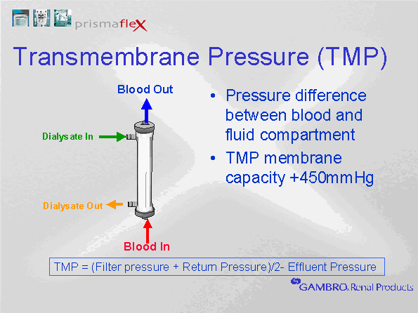
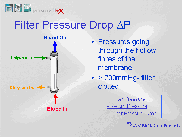
TRANSMEMBRANE PRESSURE (TMP)
The Transmembrane pressure is the hydrostatic pressure gradient across the dialysis membrane (between the blood and effluent paths). It will rise gradually at stable blood flow and effluent rates throughout the life of the filter, beginning to rise when the filter becomes plugged with clot or protein buildup. If the TMP rises suddenly, check for a kink or clamp in any of the effluent lines. A clot in the circuit will cause the TMP to rise. A high TMP with normal return pressure indicates that a filter problem (e.g., clotting). rise in as will the accumulation of clot or protein on the membrane surface.
TRANSMEMBRANE PRESSURE (TMP) =
Filter Pressure (PF) + Return Pressure (PR) - Effluent Pressure (PE)
2
TMP will increase with any increase in blood flow (an increase in blood flow increase return pressures). Stable pressures at constant blood flow rates should be expected. If the TMP increases, consider whether this occurred as a result of a new change in any flow rates.
The TMP plus the rate of TMP rise contributes to the “filter clotting” alarm. A TMP above 350 mmHg will produce an advisory alarm, and 450 mmHg will produce a "TMP excessive" alarm. The TMP and the rate of rise in the TMP contribute to the “Filter is Clotting" alarm.
DELTA P (change in filter drop)
Filter Pressure drop is another indicator of clotting. It is the difference between Filter and Return Pressure. Delta (triangle) is the symbol for change; Delta P is the rate of change in the filter drop. It will slowly rise during normal treatment.
|
|
12.
|
DIFFUSION
Small molecular weight solutes are easily removed by diffusion (dialysis). The higher the concentration gradient, the higher the diffusion rate. Solutes will move across a semipermeable membrane until the two solute concentrations become equal.
As solutes move into the dialysate fluid, the dialysate concentration of the solutes increase, reducing the diffusion gradient. Once the dialysate concentration of a solute becomes equal to the blood concentration, diffusion stops. To maintain a high diffusion gradient, the difference between the blood and dialysate concentrations must be maintained. Clearance can be increased by higher dialysate or blood flow rates. Increasing the dialysate rate maintains a low concentration of solutes on the dialysate side by increasing their removal from the dialysate fluid. Increasing the blood flow rate brings more solutes to the filter, promoting continuous diffusion. The smaller the molecule, the greater the clearance by dialysate/blood flow increases.
Although higher blood flow rates will increase the rate of clearance, CRRT circuits have limitations. The smaller filter size (compared to hemodialysis circuits) limits the blood flow rates. Blood flows can be increased substantially with hemodialysis, however, blood flow rate adjustments are limited with CRRT.
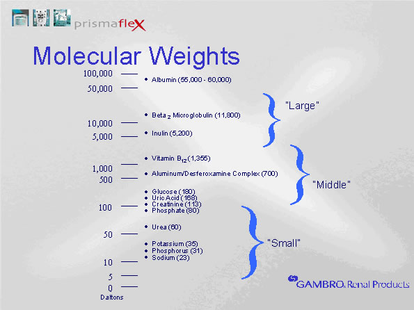
While increased dialysate flow rates enhance the clearance of small molecules, middle sized molecule clearance is more dependent upon the size of the filter pores. The only way to increase the clearance of middle sized molecules is to add convection (hemofiltration).
Optimal solute clearance is produced when dialysate flow rates are approximately double that of the blood flow rates. CRRT blood flow rates are typically 150 ml/min. A dialysate flow rate of 1 L per hour, provides a dialysate flow of 16 ml/min. Increasing the dialysate flow will have a greater effect than any increase in blood flow rates with CRRT.
Dialysate flows countercurrent, or in the opposite direction to blood flow. This promotes continual clearance by ensuring an adequate diffusion gradient is maintained. Dialysate fluid is introduced at the return end of the filter, where the serum concentration of solutes has begun to fall (due to removal from the blood within the filter). The dialysate fluid flows towards the access end of the filter where the fluid drainage tubing is located. Diffusion of solutes along the filter makes the concentration of wastes highest in the dialysate at the access end of the filter. At the access end, the blood concentration of the solute is highest , counterbalancing the rising dialysate concentration.
|
| |
HEMOFILTRATION
Dialysis effectively removes small (e.g. electrolytes) and mid size molecular weight solutes (e.g. glucose, urea, creatinine). Clearance using dialysis is determined by the solute concentration gradient and the blood flow and dialysate rates. The membrane pore size limits the ability to diffuse middle sized molecules.
One way to increase the clearance of all small and some mid sized molecules is to pull large quantities of water across the semi-permeable membrane, “dragging” additional solutes along by convection (including urea and creatinine). Hemofiltration is the removal of very large volumes of water for the purpose of increased clearance (volumes larger than desired than for fluid balance), with subsequent replacement of using an equal volume of an electrolyte solution that contains normal serum electrolyte concentratons.
Hemofiltration is being provided any time there is fluid infusing on the PreBlood Pump (predilution replacement) or on the Replacement pump (whether pre or post dilution replacement). The flow rate set on either of these two pumps will automatically be matched by an equal volume of fluid removal (replacement in = replacement out for both pumps).
Higher hemofiltration rates are of interest in critical care. Higher pre dilution rates may be a successful alternative to anticoagulant therapy. We have had success in CCTC using predilution flow rates of 2 Liters per hour. When combined with 250 - 300 ml/minute blood flow rates and good catheter flow rates, anticoagulation is often unnecessary. Although an early study by Ronco suggested that flow rates of 35 ml/kg/hr might provide enhanced cytokine clearance in sepsis, benefit has not been replicated in subsequent studies.
|
| |
THERAPIES
Original continuous hemodialysis circuits required arterial to venous access sites, because they did not utilize a blood pump to pull blood through the filter. Consequently, they were referred to as CAV (Continuous arterial-venous) circuits. Today's technology uses a blood flow pump, therefore, most continuous circuits are CVV (continuous venous-venous).
SCUF (Slow Continuous Ultrafiltration):
SCUF is the removal of water from the patient's blood as it travels through the filter. Water removal is referred to as ultrafiltration. SCUF is a therapy designed to only remove surplus water. The amount of water removed is not sufficient to remove wastes.
CVVH (Continuous Venous-Venous Hemofiltration)
CVVH is the removal of large amounts of water across the filter membrane for the purpose of clearing wastes. When large volumes of water are washed across the membrane, solutes are dragged along with the water (convection). Hemofiltration is the removal of water over and above the surplus water removed during ultrafiltration. To prevent hypovolemia, water removed during hemofiltration must be given back before the blood is returned to the patient. This is referred to as replacement. CVVH is the use of replacement fluid without dialysis fluid, plus or minus fluid removal.
CVVHD (Continuous Venous-Venous Hemodialysis):
CVVHD is the infusion of dialysis fluid into the filter canister The dialysis fluid (dialysate) surrounds the blood filled filter segments. Solutes that are small enough to fit through the membrane of the dialysis filter will move from an area of high concentration to low concentration (diffusion). The dialysate determines the solutes that will be removed. If we want to remove solutes, the concentration in the dialysate is lower than the blood concentration. If we want to give something to the patient, the concentration in the dialysate is higher than the blood. CVVHD is the removal of wastes by diffusion only, without the use of hemofiltration (replacement fluid). It can be administered with or without fluid removal from the patient.
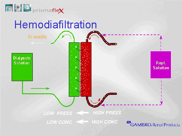
CVVHDF (Continuous Venous-Venous HemoDiaFiltration):
CVVHDF is the use of dialysis AND hemofiltration. Therapy will include the use of both dialysate and replacement fluids and can be administered with or without fluid removal from the patient.
Return to top
|
| |
REFERENCES
- Claure-Del Grandado, R. Role of acute dialysis (CRRT, SLED, Intermittent Hemodialysis). Nephrology Hypertension. https://www.renalandurologynews.com/home/decision-support-in-medicine/nephrology-hypertension/role-of-acute-dialysis-crrt-sled-intermittent-hemodialysis-other/
- Monarda, B., Rimmelea, T., Ronco, C. Extracorporeal Blood Purification Therapies for Sepsis. Blood Purif 2019;47(suppl 3):1–14
- Ronco, C. Evolution of synthetic membranes for blood purification: the case of the polyflux family. Nephrol Dial Transplant (2003) 18 [Suppl 7]: vii10–vii20
- Shafi, T. Hemodialysis: Prescription and Assessment of Adequacy.
https://www.renalandurologynews.com/home/decision-support-in-medicine/nephrology-hypertension/hemodialysis-prescription-and-assessment-of-adequacy/
- Tandukar, S., Palevsky, P. Continuous renal replacement Therapy: who, when, why and how. CHEST 2019; 155(3):626-638
- Yatzey, Al. Physical and chemical characteristics of dialysis membranes https://derangedphysiology.com/main/cicm-primary-exam/required-reading/renal-system/dialysis-and-plasmapheresis/Chapter%201165/physical-and-chemical-characteristics-dialysis-membranes#:~:text=Anatomy%20of%20the%20dialysis%20filter&text=The%20key%20features%20of%20this,and%20out%20of%20the%20filter
- Yatzey, Al. Definitions of CRRT terminology.
https://derangedphysiology.com/main/required-reading/renal-failure-and-dialysis/Chapter%203.1.1/definitions-crrt-terminology#:~:text=The%20sieving%20coefficient%20describes%20the,on%20the%20rate%20of%20ultrafiltration.&text=concentration%20%2F%20Blood%20concentration-,Relevance%20to%20CRRT%3A,of%20its%20removal%20by%20CVVHDF
- Gambro Training Manual 1 and 2
- Slides from Gambro Training package, reproduced with permission
Last Update: December 14, 2020
Last Reviewed: July 2, 2013, January 30, 2015, November 6, 2018
|
|