STANDARD OF CARE CONTINUOUS ECG MONITORING
|
Ensure that patient and health care provider safety standards are met during this procedure including:
- Risk assessment and appropriate PPE
- 4 Moments of Hand Hygiene
- Procedural Safety Pause is performed
- Two patient identification
- Safe patient handling practices
- Biomedical waste disposal policies
- Electrical safety
|
- Monitor ECG
- Maintain ECG Alarms
- Monitor ECG Rhythm Strips
|
|
STANDARD OF NURSING CARE
|
|
1.
|
Monitor ECG
All CCTC patients have continuous ECG monitoring, unless otherwise ordered. Change ECG electrodes OD and during bath, and prn.
ECG1 (the first waveform display) should be setup with the tallest (highest positive or deepest negative) QRS for optimal heart rate monitoring.
|
|
2.
|
Maintain ECG Alarms
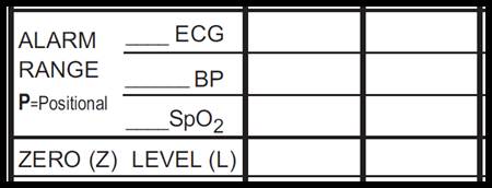
ECG alarms must be appropriately set and turned on at all times. High and low alarm settings are assessed and documented each hour in the graphic record.
Alarm settings are selected based on the degree of fluctuation in the patient's HR. Upper and lower alarm limits that represent clinically important changes are selected for each individual patient by the critical care nurse.
All alarms should be disabled during withdrawal-of-life-support. On occasion, efforts to resolve nuisance alarms may not be successful. If ECG alarms are disabled, documentation in the AI record is required. Documentation should include the reason for disabling the alarm and troubleshooting strategies.
If HR monitoring from the ECG is not possible, change the monitor to enable heart rate monitoring from the arterial line.
Confirm and alarm limits and document graphic record as shown below.
Troubleshooting
To eliminate nuissance arrhythmia alarms, turn the arrhythmia alarms to "severe". This will limit the arrhythmia detection to lethal arrhythmias. If frequent PVCs are being detected, turn the PVC alarm off.
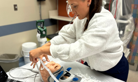
Prep skin by washing with water and drying well. Trim hair.
Do not shave the skin or use alcohol or detergents. Roughen skin up with sandpaper prior to electrode placement to improve skin contact (available in tape dispensers). Connect lead to electrode before it is applied to the skin (avoid pressing electrode onto skin; this may cause loss of contact gel).
The QRS height must be 1/2 the height of the white calibration marker. Increasing the gain WILL NOT improve the monitors ability to detect the QRS. If the patient has a low QRS voltage, look at the 12-lead ECG view and select the lead with the tallest QRS.
Moving the leads closer together, or placement of an anterior and posterior lead may increase the QRS height.
|
| 3. |
Analyze ECG Rhythm Strips
An ECG rhythm strip is collected, analyzed and posted in the clinical record at the time of admission, at the start of each shift, q4h if arrhythmias are present, q6h if cardiac status is stable and prn for significant changes in the ECG rhythm for all acute patients.
HR documentation frequency may be decreased to q shift and prn when patients become hemodynamically stable. Chronic patients with continuous ECG monitoring and stable rhythms require OD and prn ECG rhythm documentation.
|
|
|
|
|
Last Update: February 1, 2020
|