Question
of the Week:
en is a LAP (left
|
|
Answer:
The brainstem, cerebellum or occipital lobe.
Description:
The brain receives
arterial blood from the aorta via 4 vessels (2 carotid and 2 vertebral).
The right and left common carotid arteries carry blood to the middle and
anterior cerebrum. The two vertebral arteries extend from the left
and right subclavian arteries as shown in diagram 1.
 |
Diagram
1
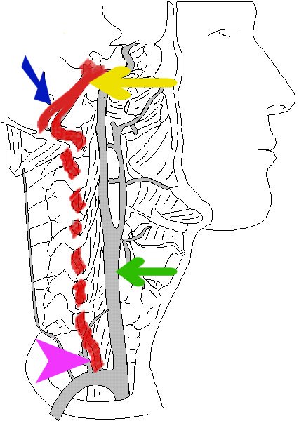
The right vertebral artery is shown in red as it leaves the right subclavian artery (pink arrow). The left vertebral artery travels up the left side and merges with the right vertebral artery (blue arrow). The right and left vertebral arteries merge to one common artery called the basilar artery (yellow arrow). The right common
carotid artery (green arrow) extends upward from the innominate artery.
|
The left and right vertebral arteries merge at the posterior base of the brain to become the basilar artery. Branches from the basilar artery supply the cerebellum, posterior cerebrum and brainstem (diagram 2).
The pontine branches
provide blood flow to the brainstem, which controls our vital centres for
breathing and the autonomic nervous system. Damage to the pons area
can cause a loss of the sympathetic control of the pupils (the sympathetic
response of the pupils is dilation), resulting in pinpoint fixed pupils.
Infarction to this area of the brain can prevent the transmission of messages
to and from the brain. This is referred to as "Locked-in" syndrome...the
patient is awake, but unable to respond (similar to a very high quadriplegia).
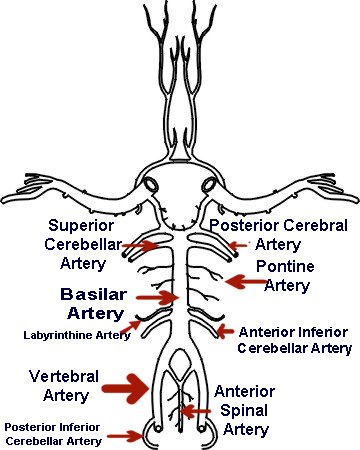 |
The posterior
cerebral artery supplies the posterior cerebrum (occipital lobe - vision),
the cerebellar branches supply the cerebellum (balance, coordination of
movements) and the pontine branches supply the brainstem. The anterior
spinal artery supplies the front of the spinal cord. The posterior
spinal artery (not shown) extends from the back of the vertebral artery
just below the anterior spinal artery bifurcation. The posterior
cerebrum, cerebellum and brainstem is shown by the arrows, below the light
blue line in diagram 3.
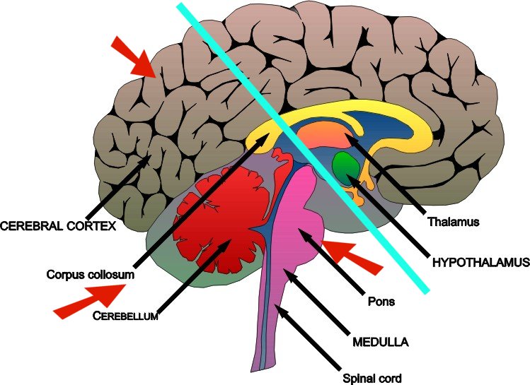 |
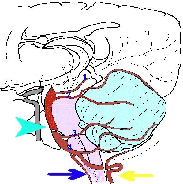
Diagram 4 shows a lateral view of the left vertebral artery (yellow arrow). The anterior spinal artery is identified by the dark blue arrow, and the basilar artery by the light blue arrow. The posterior cerebral artery (labeled 1) is shown as it extends towards the posterior cerebrum. The left occipital lobe is supplied by the left posterior cerebral artery and interprets visual stimuli from the right visual field of both eyes (e.g. images to the temporal field of the right eye and the nasal field of the left eye) .
The superior cerebellar
artery (labeled 2), the anterior inferior cerebellar artery (labeled 3)
and the posterior inferior cerebellar artery (labeled 4) collectively
provide blood flow to the cerebellum. Injury to the left hemisphere
of the cerebellum causes impaired coordination of left sided movements
(the right cerebral hemisphere "tells" the left side to move, but
the left side of the cerebellum makes the left sided movement smooth and
coordinated.
 |
Brenda Morgan:
December 17, 1990
References:
Diamond,
M., Scheibel, A., and Elson, L. (1985). The Human Brain Coloring
Book. HarperPerennial: New York. p 9.2.
|
|